Mouse EphA1 Antibody
R&D Systems, part of Bio-Techne | Catalog # AF3034

Key Product Details
Species Reactivity
Applications
Label
Antibody Source
Product Specifications
Immunogen
Glu27-Glu548
Accession # AAH71215
Specificity
Clonality
Host
Isotype
Scientific Data Images for Mouse EphA1 Antibody
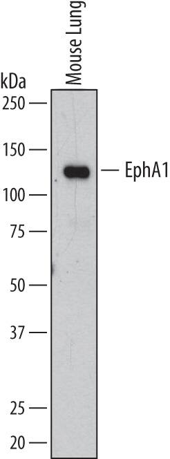
Detection of Mouse EphA1 by Western Blot.
Western blot shows lysates of mouse lung tissue. PVDF membrane was probed with 0.25 µg/mL of Goat Anti-Mouse EphA1 Antigen Affinity-purified Polyclonal Antibody (Catalog # AF3034) followed by HRP-conjugated Anti-Goat IgG Secondary Antibody (Catalog # HAF109). A specific band was detected for EphA1 at approximately 113 kDa (as indicated). This experiment was conducted under reducing conditions and using Immunoblot Buffer Group 1.EphA1 in Mouse Thymus.
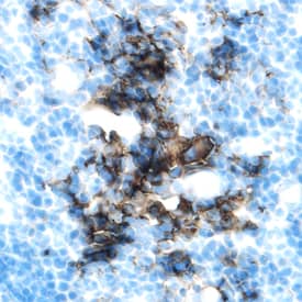
EphA1 was detected in perfusion fixed frozen sections of mouse thymus using Goat Anti-Mouse EphA1 Antigen Affinity-purified Polyclonal Antibody (Catalog # AF3034) at 1.7 µg/mL overnight at 4 °C. Tissue was stained using the Anti-Goat HRP-DAB Cell & Tissue Staining Kit (brown; Catalog # CTS008) and counterstained with hematoxylin (blue). Specific labeling was localized to thymic stromal cells. View our protocol for Chromogenic IHC Staining of Frozen Tissue Sections.Applications for Mouse EphA1 Antibody
CyTOF-ready
Flow Cytometry
Sample: D3 mouse embryonic stem cell line
Immunohistochemistry
Sample: Perfusion fixed frozen sections of mouse thymus
Western Blot
Sample: Mouse lung tissue
Formulation, Preparation, and Storage
Purification
Reconstitution
Formulation
Shipping
Stability & Storage
- 12 months from date of receipt, -20 to -70 °C as supplied.
- 1 month, 2 to 8 °C under sterile conditions after reconstitution.
- 6 months, -20 to -70 °C under sterile conditions after reconstitution.
Background: EphA1
EphA1, also known as Eph and Esk, is a member of the Eph receptor tyrosine kinase family and binds several Ephrin-A ligands. The A and B class Eph proteins share a common structural organization (1-4). The mouse EphA1 cDNA encodes a 977 amino acid (aa) precursor that includes a 24 aa signal sequence, a 524 aa extracellular domain (ECD), a 21 aa transmembrane segment, and a 408 aa cytoplasmic domain. The ECD contains an N-terminal globular domain, a cysteine-rich domain, and two fibronectin type III domains. The cytoplasmic domain contains a juxtamembrane motif with two tyrosine residues which are the major autophosphorylation sites, a kinase domain, and a conserved sterile alpha motif (SAM) (5, 6). Within the ECD, mouse EphA1 shares 84% aa sequence identity with human EphA1, approximately 40% aa sequence identity with mouse EphA2, 3, 4, 6, 7, and 8, and 27% aa sequence identity with mouse EphA5. A splice variant of mouse EphA1 lacks the transmembrane segment and is predicted to exist as a soluble molecule (7). Membrane bound or clustered Ephrin ligands interact with EphA1 and activate its kinase domain which is capable of Ser, Thr, and Tyr phosphorylation (7). Reverse signaling is propagated through the Ephrin ligand. EphA1 is widely expressed in differentiated epithelial cells, particularly in bone marrow, spleen, thymus, and testes (5, 7). It is expressed on CD4+ T cells but not on CD19+ B cells (8). On T cells, EphA1 induces Tyr phosphorylation of PYK2 and promotes chemokine-induced chemotaxis (8). EphA1 is upregulated or downregulated in a variety of human carcinomas and is implicated in tumor invasiveness (2, 9, 10).
References
- Poliakov, A. et al. (2004) Dev. Cell 7:465.
- Surawska, H. et al. (2004) Cytokine Growth Factor Rev. 15:419.
- Pasquale, E.B. (2005) Nat. Rev. Mol. Cell Biol. 6:462.
- Davy, A. and P. Soriano (2005) Dev. Dyn. 232:1.
- Coulthard, M.G. et al. (2001) Growth Factors 18:303.
- Lickliter, J.D. et al. (1996) Proc. Natl. Acad. Sci. USA 93:145.
- Douville, E.M.J. et al. (1992) Mol. Cell. Biol. 12:2681.
- Aasheim, H-C. et al. (2005) Blood 105:2869.
- Iwase, T. et al. (1993) Biochim. Biophys. Res. Commun. 194:698.
- Hirai, H. et al. (1987) Science 238:1717.
Alternate Names
Gene Symbol
UniProt
Additional EphA1 Products
Product Documents for Mouse EphA1 Antibody
Product Specific Notices for Mouse EphA1 Antibody
For research use only

