Mouse Fas/TNFRSF6/CD95 Antibody
R&D Systems, part of Bio-Techne | Catalog # AF435


Key Product Details
Species Reactivity
Validated:
Cited:
Applications
Validated:
Cited:
Label
Antibody Source
Product Specifications
Immunogen
Gly14-Arg169
Accession # P25446
Specificity
Clonality
Host
Isotype
Scientific Data Images for Mouse Fas/TNFRSF6/CD95 Antibody
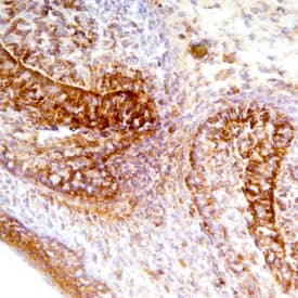
Fas/TNFRSF6/CD95 in Mouse Embryo.
Fas/TNFRSF6/CD95 was detected in immersion fixed frozen sections of mouse embryo (E15) using Goat Anti-Mouse Fas/TNFRSF6/CD95 Antigen Affinity-purified Polyclonal Antibody (Catalog # AF435) at 10 µg/mL overnight at 4 °C. Tissue was stained using the Anti-Goat HRP-DAB Cell & Tissue Staining Kit (brown; Catalog # CTS008) and counterstained with hematoxylin (blue). Specific staining was localized to olfactory epithelial cells. View our protocol for Chromogenic IHC Staining of Frozen Tissue Sections.Applications for Mouse Fas/TNFRSF6/CD95 Antibody
CyTOF-ready
Flow Cytometry
Sample: Mouse splenocytes
Immunohistochemistry
Sample: Immersion fixed frozen sections of mouse embryo (E15)
Western Blot
Sample: Recombinant Mouse Fas/TNFRSF6/CD95 Fc Chimera (Catalog # 435-FA)
Mouse Fas/TNFRSF6/CD95 Sandwich Immunoassay
Formulation, Preparation, and Storage
Purification
Reconstitution
Formulation
Shipping
Stability & Storage
- 12 months from date of receipt, -20 to -70 °C as supplied.
- 1 month, 2 to 8 °C under sterile conditions after reconstitution.
- 6 months, -20 to -70 °C under sterile conditions after reconstitution.
Background: Fas/TNFRSF6/CD95
Fas, also known as APO-1, CD95, and TNFRSF6, was originally identified as a cell-surface protein which binds to monoclonal antibodies that were cytolytic for various human cell lines. In the TNF receptor superfamily nomenclature, Fas is referred to as TNFRSF6. Human and mouse Fas cDNAs encode a 325 and a 327 amino acid residue type 1 membrane protein, respectively, that belongs to the TNF and NGF receptor family. Alternatively spliced cDNAs encoding multiple human Fas isoforms, including a soluble form of Fas lacking the transmembrane domain, have also been identified. Fas is highly expressed in epithelial cells, hepatocytes, activated mature lymphocytes, virus-transformed lymphocytes and other tumor cells. Fas expression has also been detected in mouse thymus, liver, heart, lung, kidney and ovary. The ligand for Fas (FasL) has been identified and shown to be a member of the TNF family of type 2 membrane proteins. FasL is predominantly expressed by activated T-lymphocytes, NK cells, and in tissues with immune-privileged sites. Soluble FasL can be produced by proteolysis of membrane-associated Fas.
Ligation of Fas by FasL or anti-Fas antibody has been shown to induce apoptotic cell death in Fas-bearing cells. Fas plays a role in the down-regulation of the immune reaction and has been shown to be a key mediator of activation-induced death of activated T lymphocytes. Fas-mediated cell death has also been shown to be important for the deletion of activated or autoreactive B lymphocytes. Besides the perforin/granzyme-based mechanism, the Fas system has been identified as the alternate pathway for CTL-mediated cytotoxicity. FasL has also been shown to function in immunological privileged sites by killing infiltrating Fas-bearing lymphocytes and inflammatory cells.
References
- Nagata, S. and P. Golstein (1995) Science 267:1449.
- Nagata, S. (1997) Cell 88:355.
- Parijs, L. and A.K. Abbas (1996) Current Opinion in Immunol. 8:355.
- Green, D.R. and C.F. Ware (1997) Proc. Natl. Acad. Sci. USA. 94:5986.
Long Name
Alternate Names
Entrez Gene IDs
Gene Symbol
UniProt
Additional Fas/TNFRSF6/CD95 Products
Product Documents for Mouse Fas/TNFRSF6/CD95 Antibody
Product Specific Notices for Mouse Fas/TNFRSF6/CD95 Antibody
For research use only