Mouse Galectin-7 Antibody
R&D Systems, part of Bio-Techne | Catalog # MAB1304

Key Product Details
Species Reactivity
Applications
Label
Antibody Source
Product Specifications
Immunogen
Ser2-Phe136
Accession # AAK29385
Specificity
Clonality
Host
Isotype
Scientific Data Images for Mouse Galectin-7 Antibody
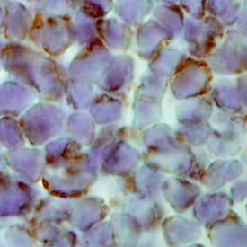
Galectin‑7 in Mouse Thymus.
Galectin-7 was detected in perfusion fixed frozen sections of mouse thymus using Rat Anti-Mouse Galectin-7 Monoclonal Antibody (Catalog # MAB1304) at 25 µg/mL overnight at 4 °C. Tissue was stained using the Anti-Rat HRP-DAB Cell & Tissue Staining Kit (brown; Catalog # CTS017) and counterstained with hematoxylin (blue). Specific labeling was localized to the cytoplasm of lymphocytes. View our protocol for Chromogenic IHC Staining of Frozen Tissue Sections.Applications for Mouse Galectin-7 Antibody
Immunohistochemistry
Sample: Perfusion fixed frozen sections of mouse skin and thymus
Western Blot
Sample: Recombinant Mouse Galectin-7 (Catalog # 1304-GA)
Mouse Galectin-7 Sandwich Immunoassay
Reviewed Applications
Read 1 review rated 5 using MAB1304 in the following applications:
Formulation, Preparation, and Storage
Purification
Reconstitution
Formulation
Shipping
Stability & Storage
- 12 months from date of receipt, -20 to -70 °C as supplied.
- 1 month, 2 to 8 °C under sterile conditions after reconstitution.
- 6 months, -20 to -70 °C under sterile conditions after reconstitution.
Background: Galectin-7
The galectins constitute a large family of carbohydrate-binding proteins with specificity for N-acetyl-lactosamine-containing glycoproteins. At least 14 mammalian galectins, which share structural similarities in their carbohydrate recognition domains (CRD), have been identified. The galectins have been classified into the prototype galectins (-1, -2, -5, -7, -10, -11, -13, -14), which contain one CRD and exist either as a monomer or a noncovalent homodimer; the chimera galectins (Galectin-3) containing one CRD linked to a nonlectin domain; and the tandem-repeat galectins (-4, -6, -8, -9, -12) consisting of two CRDs joined by a linker peptide. Galectins lack a classical signal peptide and can be localized to the cytosolic compartments where they have intracellular functions. However, via one or more as yet unidentified non-classical secretory pathways, galectins can also be secreted to function extracellularly. Individual members of the galectin family have different tissue distribution profiles and exhibit subtle differences in their carbohydrate-binding specificities. Each family member may preferentially bind to a unique subset of cell-surface glycoproteins (1‑4).
Mouse Galectin-7 is a prototype monomeric galectin. It is expressed in stratified epithelia and is significantly down-regulated in squamous cell carcinomas. Galectin-7 is a pro-apoptotic protein that is highly induced by the tumor suppressor protein p53. It functions intracellularly upstream of JNK activation to enhance cytochrome c release during apoptosis (5). Galectin-7 may also be involved in cell-cell and cell-matrix interactions and exogenous galectin has been found to accelerate the re‑epithelialization of wounds (6).
References
- Rabinovich, A. et al. (2002) TRENDS in Immunol. 23:313.
- Rabinovich, A. et al. (2002) J. Leukocyte Biology 71:741.
- Hughes, R.C. (2002) Biochimie 83:667.
- R&D Systems' Cytokine Bulletin, Summer (2002).
- Kuwabara, I. et al. (2002) J. Biol. Chem. 277:3487.
- Cao, Z. et al. (2002) J. Biol. Chem. 277:42299.
Alternate Names
Gene Symbol
UniProt
Additional Galectin-7 Products
Product Documents for Mouse Galectin-7 Antibody
Product Specific Notices for Mouse Galectin-7 Antibody
For research use only
