Mouse H60 PE-conjugated Antibody
R&D Systems, part of Bio-Techne | Catalog # FAB1155P


Key Product Details
Species Reactivity
Validated:
Cited:
Applications
Validated:
Cited:
Label
Antibody Source
Product Specifications
Immunogen
Asp30-Gln212
Accession # Q3TDZ7
Specificity
Clonality
Host
Isotype
Scientific Data Images for Mouse H60 PE-conjugated Antibody
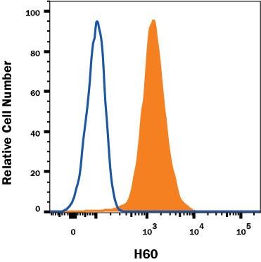
Detection of H60 in RAW 264.7 Mouse Cell Line by Flow Cytometry.
RAW 264.7 mouse monocyte/macrophage cell line was stained with Rat Anti-Mouse H60 PE-conjugated Monoclonal Antibody (Catalog # FAB1155P, filled histogram) or isotype control antibody (Catalog # IC006P, open histogram). View our protocol for Staining Membrane-associated Proteins.Applications for Mouse H60 PE-conjugated Antibody
Flow Cytometry
Sample: RAW 264.7 mouse monocyte/macrophage cell line
Formulation, Preparation, and Storage
Purification
Formulation
Shipping
Stability & Storage
- 12 months from date of receipt, 2 to 8 °C as supplied.
Background: H60
H60 was originally described as an immunodominant histocompatibility antigen that is expressed in BALB mice but not in B6 mice. More recently it was reported to function as a ligand for mouse NKG2D, an activating receptor found on NK cells, on some T cell subsets, and on stimulated macrophages. H60 shares approximately 25 percent amino acid identity with the Rae-1 family, a small group of proteins that also function as ligands for mouse NKG2D. H60 and the Rae-1 proteins are distantly related to MHC class I proteins, but they possesses only the alpha1 and alpha2 Ig-like domains, and have no capacity to bind peptide or interact with beta2-microglobulin. The genes encoding these proteins are not found within the Major Histocompatibility Complex on mouse chromosome 17, but rather map to mouse chromosome 10. Unlike the GPI-linked Rae-1 proteins, H60 appears to be anchored to the membrane via a hydrophobic transmembrane segment. H60 transcripts were found in embryonic tissue, in spleen, and in some transformed cell lines. Transcripts were also observed in mouse skin cells after exposure to carcinogens. Binding of H60 to NKG2D results in the activation of cytolytic activity and/or cytokine production by the NKG2D-expressing effector cells. Ectopic expression of H60 on mouse tumor cell lines resulted in the in vivo rejection of the tumors (1-6).
References
- Malarkannan, S. et al. (1998) J. Immunol. 161:3501.
- Diefenbach, A. et al. (2000) Nature Immunol. 1:119.
- Cerwenka, A. et al. (2000) Immunity 12:721.
- Cerwenka, A. et al. (2001) Proc. Natl. Acad. Sci. USA 98:11521.
- Diefenbach, A. et al. (2001) Nature 413:165.
- NKG2D and its Ligands, www.RnDSystems.com.
Long Name
Alternate Names
Entrez Gene IDs
Gene Symbol
UniProt
Additional H60 Products
Product Documents for Mouse H60 PE-conjugated Antibody
Product Specific Notices for Mouse H60 PE-conjugated Antibody
For research use only