Mouse IL-22 Antibody
R&D Systems, part of Bio-Techne | Catalog # AF582


Key Product Details
Species Reactivity
Validated:
Cited:
Applications
Validated:
Cited:
Label
Antibody Source
Product Specifications
Immunogen
Leu34-Val179
Accession # Q9JJY9
Specificity
Clonality
Host
Isotype
Endotoxin Level
Scientific Data Images for Mouse IL-22 Antibody
IL‑10 Secretion Induced by IL‑22 and Neutralization by Mouse IL‑22 Antibody.
Recombinant Mouse IL-22 (Catalog # 582-ML) stimulates IL-10 secretion in the COLO 205 human colorectal adenocarcinoma cell line in a dose-dependent manner (orange line), as measured by the Human IL-10 DuoSet ELISA Development Kit (Catalog # DY217B). IL-10 secretion elicited by Recombinant Mouse IL-22 (1 ng/mL) is neutralized (green line) by increasing concentrations of Goat Anti-Mouse IL-22 Antigen Affinity-purified Polyclonal Antibody (Catalog # AF582). The ND50 is typically 0.5-2.5 µg/mL.Detection of Mouse Mouse IL-22 Antibody by Flow Cytometry
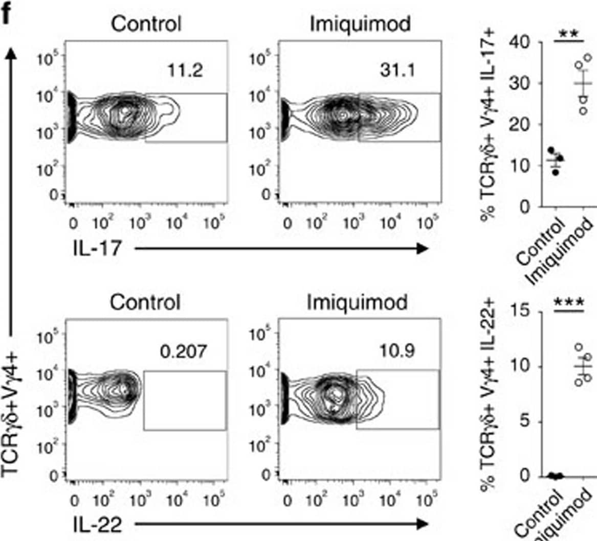
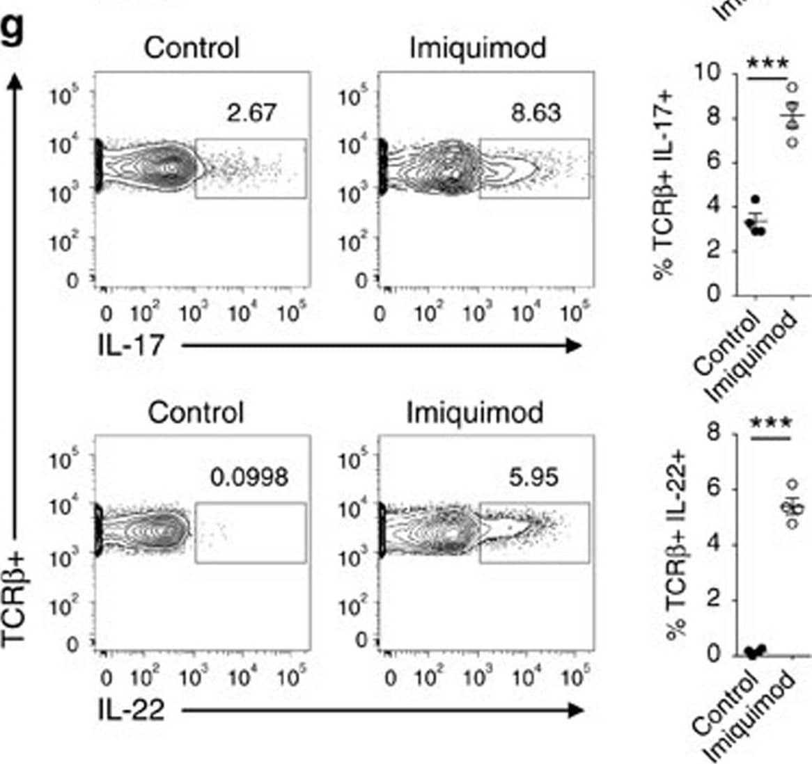
Imiquimod increases IL-17 and IL-22 in both gamma delta and alpha beta T cells in skin.Skin and ear of wild type C57BL/6 mice were treated by topical application of imiquimod cream (Fougera, n=4) or control cream (Vaseline, n=3) for 6 consecutive days. (a) Weight loss of imiquimod and control cream treated mice monitored daily. (b) Photographs of imiquimod and control cream treated skin and ear; photos were taken at day 6. H&E stained ear and back skin sections of imiquimod treated and control mice. Scale bar, 200 μm. (c) Thickness of skin measured by Digimatic Caliper at day 6 in control and imiquimod treated mice. Data showed represents average of at least two measurements. (d) Ear thickness of imiquimod and control cream treated mice monitored daily. Ear thickness was measured using Digimatic Caliper. (e) Quantitative PCR analysis of Th17 associated cytokines and Foxp3 in skin after 6 days of imiquimod and control cream treatment. (f) Representative flow cytometric analysis of TCR gamma delta+V gamma4+IL-17+ (upper raw) and TCR gamma delta+V gamma4+IL-22+ cells (lower row) in the skin of control (n=3) and imiquimod treated mice (n=4). (g) Representative flow cytometric analysis of single-positive TCR beta+IL-17+ (upper raw) or TCR beta+IL-22+ (lower raw) cells in the skin of control (n=3) and imiquimod treated mice (n=4). Data representative of more than three experiments, results are shown as mean±s.e.m., significance determined by unpaired two-tailed Student's t-test (*P<0.05; **P<0.01; ***P<0.001). Image collected and cropped by CiteAb from the following publication (https://pubmed.ncbi.nlm.nih.gov/26416167), licensed under a CC-BY license. Not internally tested by R&D Systems.Detection of Mouse Mouse IL-22 Antibody by Flow Cytometry
Imiquimod increases IL-17 and IL-22 in both gamma delta and alpha beta T cells in skin.Skin and ear of wild type C57BL/6 mice were treated by topical application of imiquimod cream (Fougera, n=4) or control cream (Vaseline, n=3) for 6 consecutive days. (a) Weight loss of imiquimod and control cream treated mice monitored daily. (b) Photographs of imiquimod and control cream treated skin and ear; photos were taken at day 6. H&E stained ear and back skin sections of imiquimod treated and control mice. Scale bar, 200 μm. (c) Thickness of skin measured by Digimatic Caliper at day 6 in control and imiquimod treated mice. Data showed represents average of at least two measurements. (d) Ear thickness of imiquimod and control cream treated mice monitored daily. Ear thickness was measured using Digimatic Caliper. (e) Quantitative PCR analysis of Th17 associated cytokines and Foxp3 in skin after 6 days of imiquimod and control cream treatment. (f) Representative flow cytometric analysis of TCR gamma delta+V gamma4+IL-17+ (upper raw) and TCR gamma delta+V gamma4+IL-22+ cells (lower row) in the skin of control (n=3) and imiquimod treated mice (n=4). (g) Representative flow cytometric analysis of single-positive TCR beta+IL-17+ (upper raw) or TCR beta+IL-22+ (lower raw) cells in the skin of control (n=3) and imiquimod treated mice (n=4). Data representative of more than three experiments, results are shown as mean±s.e.m., significance determined by unpaired two-tailed Student's t-test (*P<0.05; **P<0.01; ***P<0.001). Image collected and cropped by CiteAb from the following publication (https://pubmed.ncbi.nlm.nih.gov/26416167), licensed under a CC-BY license. Not internally tested by R&D Systems.Applications for Mouse IL-22 Antibody
Western Blot
Sample: Recombinant Mouse IL-22 (Catalog # 582-ML)
Neutralization
Mouse IL-22 Sandwich Immunoassay
Reviewed Applications
Read 1 review rated 4 using AF582 in the following applications:
Formulation, Preparation, and Storage
Purification
Reconstitution
Formulation
Shipping
Stability & Storage
- 12 months from date of receipt, -20 to -70 °C as supplied.
- 1 month, 2 to 8 °C under sterile conditions after reconstitution.
- 6 months, -20 to -70 °C under sterile conditions after reconstitution.
Background: IL-22
Interleukin-22 (IL-22), also known as IL-10-related T cell-derived inducible factor (IL-TIF) was initially identified as a gene induced by IL-9 in mouse T cells and mast cells. Mouse IL-22 cDNA encodes a 179 amino acid (aa) residue protein with a putative 33 aa signal peptide that is cleaved to generate a 147 aa mature protein that shares approximately 79% and 22% aa sequence identity with human IL-22 and IL-10, respectively. The mouse IL-22 gene is localized to chromosome 10. Although it exists as a single copy gene in many mouse strains, the IL-22 gene is duplicated in some mouse strains including C57B1/6, FVB and 129. The two mouse genes designated IL-TIF alpha and IL-TIF beta, share greater than 98% sequence homology in their coding region. IL-22 has been shown to activate STAT1 and STAT3 in several hepatoma cell lines and upregulate the production of acute phase proteins. IL-22 is produced by normal mouse T cells upon Con A activation. Mouse IL-22 expression is also induced in various organs upon lipopolysaccharide injection, suggesting that IL-22 may be involved in inflammatory responses. The functional IL-22 receptor complex consists of two receptor subunits, IL-22 R (previously an orphan receptor named CRF2-9) and IL-10 R beta (previously known as CRF2-4), belonging to the class II cytokine receptor family.
Long Name
Alternate Names
Gene Symbol
UniProt
Additional IL-22 Products
Product Documents for Mouse IL-22 Antibody
Product Specific Notices for Mouse IL-22 Antibody
For research use only

