Mouse IL-6R alpha Antibody
R&D Systems, part of Bio-Techne | Catalog # AF1830


Key Product Details
Species Reactivity
Validated:
Cited:
Applications
Validated:
Cited:
Label
Antibody Source
Product Specifications
Immunogen
Leu20-Glu357
Accession # P22272
Specificity
Clonality
Host
Isotype
Endotoxin Level
Scientific Data Images for Mouse IL-6R alpha Antibody
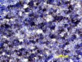
IL-6R alpha in Mouse Thymus.
IL-6R alpha was detected in perfusion fixed frozen sections of mouse thymus using Mouse IL-6R alpha Antigen Affinity-purified Polyclonal Antibody (Catalog # AF1830) at 15 µg/mL overnight at 4 °C. Tissue was stained using the Anti-Goat HRP-DAB Cell & Tissue Staining Kit (brown; Catalog # CTS008) and counterstained with hematoxylin (blue). View our protocol for Chromogenic IHC Staining of Frozen Tissue Sections.IL-6R alpha in Mouse Thymus.
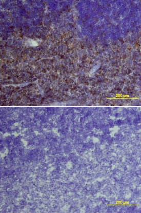
IL-6R alpha was detected in perfusion fixed frozen sections of mouse thymus using Mouse IL-6R alpha Antigen Affinity-purified Polyclonal Antibody (Catalog # AF1830) at 15 µg/mL overnight at 4 °C. Tissue was stained using the Anti-Goat HRP-DAB Cell & Tissue Staining Kit (brown; Catalog # CTS008) and counterstained with hematoxylin (blue). Lower panel shows a lack of labeling if primary antibodies are omitted and tissue is stained only with secondary antibody followed by incubation with detection reagents. View our protocol for Chromogenic IHC Staining of Frozen Tissue Sections.Cell Proliferation Induced by IL‑6 and Neutralization by Mouse IL-6R alpha Antibody.
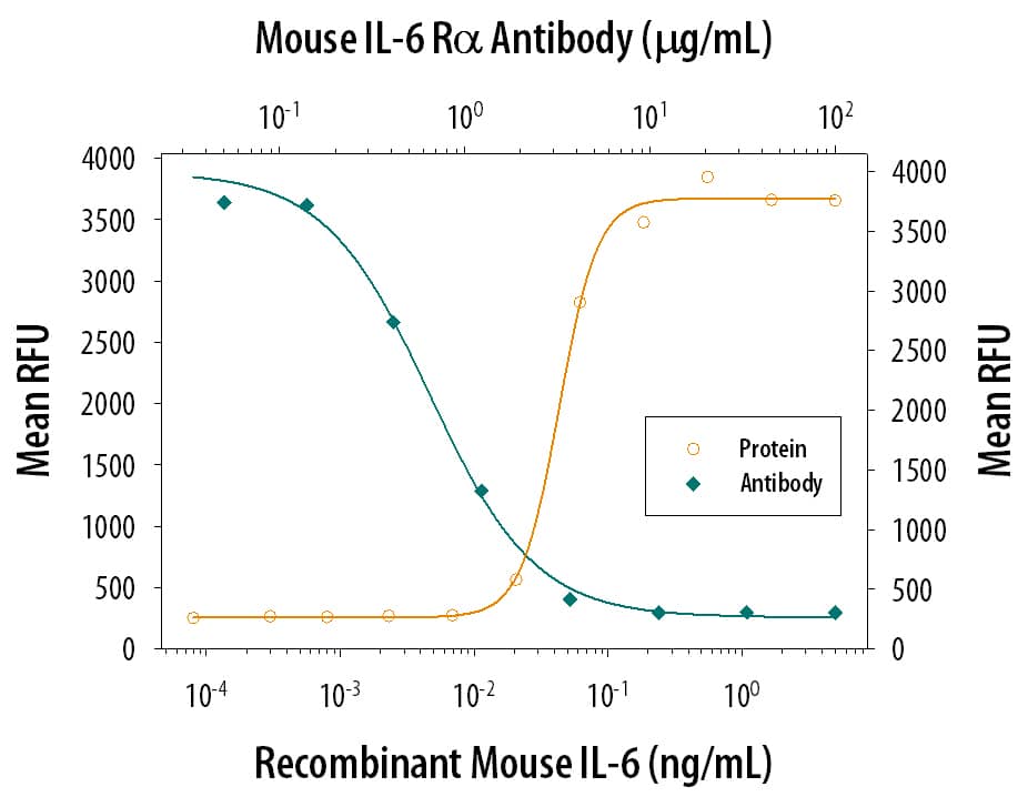
Recombinant Mouse IL-6 (Catalog # 406-ML) stimulates proliferation in the T1165.85.2.1 mouse plasmacytoma cell line in a dose-dependent manner (orange line). Proliferation elicited by Recombinant Mouse IL-6 (0.25 ng/mL) is neutralized (green line) by increasing concentrations of Mouse IL-6R alpha Antigen Affinity-purified Polyclonal Antibody (Catalog # AF1830). The ND50 is typically 0.2-1 µg/mL.Applications for Mouse IL-6R alpha Antibody
CyTOF-ready
Flow Cytometry
Sample: Mouse CD3+ splenocytes
Immunohistochemistry
Sample: Perfusion fixed frozen sections of mouse thymus
Western Blot
Sample: Recombinant Mouse IL-6R alpha (Catalog # 1830-SR)
Neutralization
Mouse IL-6R alpha Sandwich Immunoassay
Formulation, Preparation, and Storage
Purification
Reconstitution
Formulation
Shipping
Stability & Storage
- 12 months from date of receipt, -20 to -70 °C as supplied.
- 1 month, 2 to 8 °C under sterile conditions after reconstitution.
- 6 months, -20 to -70 °C under sterile conditions after reconstitution.
Background: IL-6R alpha
Interleukin 6 (IL-6) is a multifunctional cytokine that exerts its activities by binding to a high-affinity receptor complex consisting of two membrane glycoproteins: an 80 kDa ligand binding subunit (IL-6 R alpha/CD126) and a 130 kDa nonligand-binding signal-transducing subunit (gp130/CD130) (1‑4). The mouse IL-6 R alpha cDNA encodes a precursor type I transmembrane protein of 460 amino acids (aa) that contains a 19 aa signal sequence, a 345 aa extracellular ligand binding domain, a 21 aa transmembrane region, and a 75 aa cytoplasmic segment (2). The extracellular segment contains an Ig-like and a fibronectin-type III domain, plus a membrane proximal WSXWS motif. In their extracellular regions, mouse IL-6 R alpha shares 89%, 51% and 50% aa identity with rat, human and porcine IL-6 R alpha, respectively. Unlike gp130 that is expressed ubiquitously, the cellular distribution of IL-6 R alpha is predominantly limited to hepatocytes and leukocyte subpopulations such as monocytes, neutrophils, T and B cells. Soluble IL-6 R alpha has been found in various body fluids (5). Two soluble receptor isoforms that arise either from proteolytic cleavage of the membrane-bound IL-6 R alpha, or by alternative mRNA splicing (reported only in human) have been described (6, 7). Soluble IL-6 R alpha binds IL-6 with an affinity similar to that of the membrane-bound IL-6 R alpha. More importantly, the soluble IL-6 R alpha/IL-6 complex is capable of interacting with the membrane-bound gp130 to activate cells that lack an integral membrane IL-6 R alpha. It has been documented that elevated soluble IL-6 R is associated with numerous diseases including arthritic lesions, multiple myeloma and Crohn’s disease (6, 7).
References
- Yamasaki, K. et al. (1988) Science 241:825.
- Sugita, T. et al. (1990) J. Exp. Med. 171:2001.
- Hibi, M. et al. (1990) Cell 63:1149.
- Saito, M. et al. (1992) J. Immunol. 148:4066.
- Novick, D. et al. (1989) J. Exp. Med. 170:1409.
- Jones, S.A. et al. (2001) FASEB J. 15:43.
- Jones, S.A. and S. Rose-John (2002) Biochim. Biophys. Acta 1592:251.
Long Name
Alternate Names
Gene Symbol
UniProt
Additional IL-6R alpha Products
Product Documents for Mouse IL-6R alpha Antibody
Product Specific Notices for Mouse IL-6R alpha Antibody
For research use only

