Mouse Integrin alpha9 Antibody
R&D Systems, part of Bio-Techne | Catalog # AF3827


Key Product Details
Validated by
Biological Validation
Species Reactivity
Validated:
Mouse
Cited:
Human, Mouse, Transgenic Mouse
Applications
Validated:
CyTOF-ready, Flow Cytometry, Immunohistochemistry, Western Blot
Cited:
Flow Cytometry, IHC/IF, Immunocytochemistry, Immunohistochemistry, Immunohistochemistry-Frozen, Neutralization, Western Blot
Label
Unconjugated
Antibody Source
Polyclonal Goat IgG
Product Specifications
Immunogen
Mouse myeloma cell line NS0-derived recombinant mouse Integin alpha9
Try31-Val979
Accession # NP_598482
Try31-Val979
Accession # NP_598482
Specificity
Detects mouse Integrin alpha9 in direct ELISAs and Western blots. In direct ELISAs, less than 25% cross-reactivity with recombinant human Integrin alpha9 is observed and less than 5% cross-reactivity with recombinant mouse Integrin alpha3 and alpha4 is observed.
Clonality
Polyclonal
Host
Goat
Isotype
IgG
Scientific Data Images for Mouse Integrin alpha9 Antibody
Integrin alpha 9 in Mouse Liver Tissue.
Integrin a9 was detected in perfusion fixed frozen sections of mouse liver tissue using Goat Anti-Mouse Integrin a9 Antigen Affinity-purified Polyclonal Antibody (Catalog # AF3827) at 1 µg/mL overnight at 4 °C. Before incubation with the primary antibody, tissue was subjected to heat-induced epitope retrieval using Antigen Retrieval Reagent-Basic (Catalog # CTS013). Tissue was stained using the Anti-Goat IgG VisUCyte™ HRP Polymer Antibody (brown; Catalog # VC004) and counterstained with hematoxylin (blue). Specific staining was localized to bile canaliculi. View our protocol for IHC Staining with VisUCyte HRP Polymer Detection Reagents.Detection of Human Integrin alpha 9 by Immunocytochemistry/Immunofluorescence
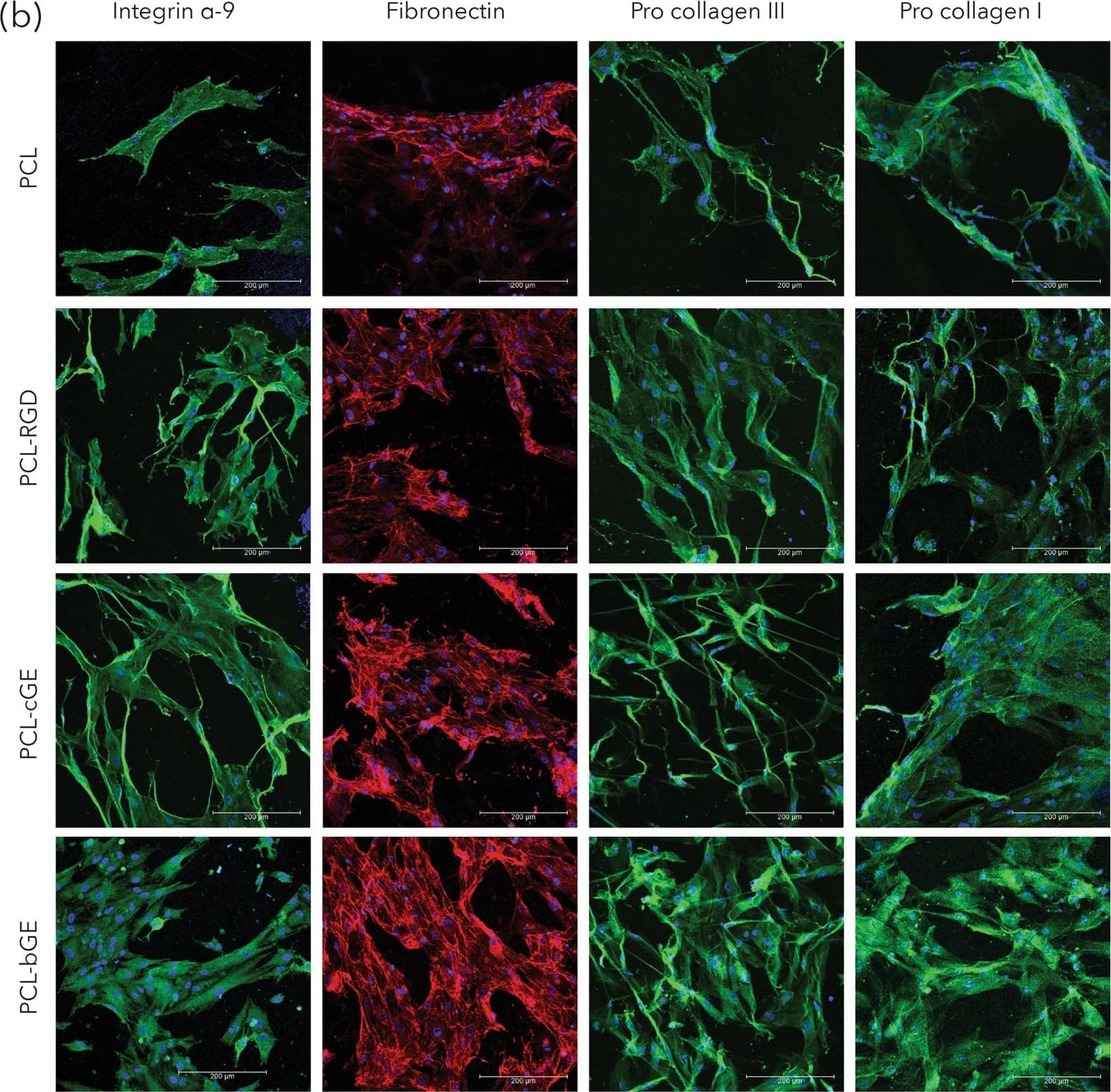
Morphology and protein expression of hSKPs. (a) SEM micrographs of nanofibrous scaffolds showing morphology of hSKPs at 7 days of culture. (b) Immunofluorescent images of hSKPs grown on nanofibrous scaffolds showing expression of integrin alpha-9 (green), fibronectin (red) and procollagen I & III (green). Image collected and cropped by CiteAb from the following publication (https://pubmed.ncbi.nlm.nih.gov/28860484), licensed under a CC-BY license. Not internally tested by R&D Systems.Detection of Mouse Integrin alpha 9 by Immunocytochemistry/Immunofluorescence
Intergins alpha4/ alpha9 are responsible for the action of osteopontin (OPN) in the augmentation of macrophage (Mφ) M2 polarization and angiogenic capacity. (A) Relative mean fluorescence intensities (MFIs) of integrins alpha4/ alpha9 were measured by FACS analysis in each Mφ subset. Anti-integrin alpha4 or alpha9 antibodies (Abs) and each isotype-matched control Ab were used at 10 µg/mL, n = 4 [***p < 0.001 vs. untreated, ###p < 0.001 vs. interleukin (IL)-10 alone]. (B) Representative confocal laser scanning immunofluorescence overlay images of integrin alpha4 (green) and DAPI (blue), as well as those of integrin alpha9 (red) and DAPI (blue) in Mφ (–) and Mφ (IL-10 + IL-18). Scale bar represents 20 µm. (C) Relative MFI of CD163 was measured by FACS analysis in each Mφ subset. Anti-integrin alpha4 or alpha9 Abs and each isotype-matched control Ab were used at 10 µg/mL, n = 4 (***p < 0.001 vs. untreated, ###p < 0.001 vs. IL-10 alone, †††p < 0.001 vs. IL-10 + IL-18). (D) The total areas and lengths of tube-like structures were determined by the Matrigel tube formation assay where b.End5 was cocultured with each Mφ subset. Anti-integrin alpha4 or alpha9 Abs and each isotype-matched control Ab were used at 10 µg/mL, n = 6 (***p < 0.001, **p < 0.01, *p < 0.05 vs. untreated, ###p < 0.001, ##p < 0.01, #p < 0.05 vs. IL-10 alone, †††p < 0.001 vs. IL-10 + IL-18). Integrin alpha4 = ITGA4; Integrin alpha9 = ITGA9. All data are expressed as means ± SEM and were analyzed by a one-way ANOVA followed by Tukey’s test. Image collected and cropped by CiteAb from the following publication (https://pubmed.ncbi.nlm.nih.gov/29559970), licensed under a CC-BY license. Not internally tested by R&D Systems.Applications for Mouse Integrin alpha9 Antibody
Application
Recommended Usage
CyTOF-ready
Ready to be labeled using established conjugation methods. No BSA or other carrier proteins that could interfere with conjugation.
Flow Cytometry
2.5 µg/106 cells
Sample: D3 mouse embryonic stem cell line
Sample: D3 mouse embryonic stem cell line
Immunohistochemistry
1-15 µg/mL
Sample: Perfusion fixed frozen sections of mouse liver
Sample: Perfusion fixed frozen sections of mouse liver
Western Blot
0.1 µg/mL
Sample: Recombinant Mouse Integrin alpha9
Sample: Recombinant Mouse Integrin alpha9
Formulation, Preparation, and Storage
Purification
Antigen Affinity-purified
Reconstitution
Reconstitute at 0.2 mg/mL in sterile PBS. For liquid material, refer to CoA for concentration.
Formulation
Lyophilized from a 0.2 μm filtered solution in PBS with Trehalose. *Small pack size (SP) is supplied either lyophilized or as a 0.2 µm filtered solution in PBS.
Shipping
Lyophilized product is shipped at ambient temperature. Liquid small pack size (-SP) is shipped with polar packs. Upon receipt, store immediately at the temperature recommended below.
Stability & Storage
Use a manual defrost freezer and avoid repeated freeze-thaw cycles.
- 12 months from date of receipt, -20 to -70 °C as supplied.
- 1 month, 2 to 8 °C under sterile conditions after reconstitution.
- 6 months, -20 to -70 °C under sterile conditions after reconstitution.
Background: Integrin alpha 9
Alternate Names
ITGA4L, ITGA9, RLC-alpha
Gene Symbol
ITGA9
UniProt
Additional Integrin alpha 9 Products
Product Documents for Mouse Integrin alpha9 Antibody
Product Specific Notices for Mouse Integrin alpha9 Antibody
For research use only
Loading...
Loading...
Loading...
Loading...
Loading...

