Mouse Netrin-1 Antibody
R&D Systems, part of Bio-Techne | Catalog # AF1109


Key Product Details
Validated by
Species Reactivity
Validated:
Cited:
Applications
Validated:
Cited:
Label
Antibody Source
Product Specifications
Immunogen
Val22-Ala603
Accession # AAC52971
Specificity
Clonality
Host
Isotype
Endotoxin Level
Scientific Data Images for Mouse Netrin-1 Antibody
Detection of Human Netrin-1 by Western Blot
UNC5A and UNC5C induce caspase-3 activation through DAPK1/PR65b during UPR. (A) Quantification of netrin-1–receptor expression by qRT-PCR (mean + SEM, n = 3). (B–F) Identification of the implicated netrin-1 receptors. HepaRG cells were transfected with siRNAs and treated with DTT or not for 4 hours (mock). (B) Assessment of XBP1 mRNA splicing by RT-PCR. Representative result, n = 3. (C) Assessment of netrin-1 protein knockdown by immunoblotting. Representative result, n = 3. (D and E) Assessment of transcript knockdown efficiencies. Graphs indicate (D) UNC5A and (E) UNC5C mRNA levels in siRNA-treated cells in comparison with control siRNA-treated cells (mean + SEM; n = 3; Mann–Whitney test; P < .05). (F) Caspase-3 activation is reversed by UNC5A or UNC5C knockdown after UPR induction. Graph indicates caspase-3 activity ratio of DTT vs untreated cells for each condition (mean + SEM; n = 3; Mann–Whitney test; P < .05). (G–J) Identification of the downstream signaling pathway. HepaRG cells were transfected with siRNAs and treated with DTT or not for 4 hours (mock). (G) Assessment of XBP1 mRNA splicing by RT-PCR. Representative result, n = 3. (H) Evaluation of netrin-1, DAPK1, and PR65 beta depletion by immunoblotting. Representative result, n = 3. (I) Caspase-3 activation is reversed by DAPK1 or PR65 beta knockdown. Graph indicates the caspase-3 activity ratio for each condition (mean + SEM; n = 3; Mann–Whitney test; P < .05). (J) PP2A activity is increased by netrin-1 depletion and reversed by reduced expression of PR65 beta. Graph indicates PP2A activity ratio for each condition (mean + SEM; n = 3; Mann–Whitney test; P < .05). *,**, or *** refer to statistical analyses. Image collected and cropped by CiteAb from the following open publication (https://pubmed.ncbi.nlm.nih.gov/28174720), licensed under a CC-BY license. Not internally tested by R&D Systems.Detection of Human Netrin-1 by Western Blot
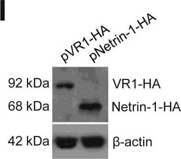
Netrin-1 protects against cell death during UPR. (A–H) Netrin-1 depletion. (A–E) HepaRG cells were transfected with netrin-1 siRNA, treated with DTT, and harvested in a time course assay. (A) Netrin-1 protein knockdown by siRNA was validated by immunoblot (representative result, n = 3). (B) XBP1 splicing was confirmed by RT-PCR (representative result, n = 3). (C) UPR increases the dependence of cells toward netrin-1 for survival. Graphs indicate the difference in cell death (in percentages) (mean + SEM; n = 3; Mann–Whitney test; P < .05). (D) UPR increases the sensitivity of cells toward netrin-1 for caspase-3 activation. Graphs indicate caspase-3 activity ratios (mean + SEM; n = 3; Mann–Whitney test; P < .05). (E) Netrin-1 depletion increases apoptosis. Graph indicates apoptotic cell death ratio by propidium iodide staining and flow cytometry (mean + SEM; n = 3; Mann–Whitney test; P < .05). (F–H) Netrin-1 inhibition using a neutralizing antibody. HepaRG cells were seeded and treated with a control (H4) or anti–netrin-1 antibody (2F5) the same day. Cells were treated with DTT 3 days after addition and harvested after the indicated time points. (F) Assessment of XBP1 mRNA splicing (representative result, n = 3). (G) Netrin-1 neutralization enhances caspase-3 activity. Graph indicates the caspase-3 activity ratios between 2F5 and H4-treated cells (mean + SEM; n = 3; Mann–Whitney test; P < .05). (H) Netrin-1 depletion increases apoptosis. Graph indicates the apoptotic cell death ratio as assessed after propidium iodide staining and flow cytometry of netrin-1–depleted and control cells (mean + SEM; n = 3; Mann–Whitney test; P < .05). (I–M) Netrin-1 forced expression. HepaRG cells were transfected with control (VR1-HA) and netrin-1 (netrin-1–HA) vectors, treated with DTT, and harvested. (I) Netrin-1 expression was assessed by immunoblotting. Representative result, n = 3. (J) XBP1 mRNA splicing was quantified by RT-PCR at the indicated time points. Representative result, n = 3. (K) UPR increases the sensitivity of cells toward netrin-1 for protection against cell death in time course assays. Graph indicates the difference in cell death (in percentages) between netrin-1–overexpressing cells and control cells (mean + SEM; n = 3; Mann–Whitney test; P < .05). (L) UPR increases the sensitivity of cells toward netrin-1 for caspase-3 activation in time course assays. Graph indicates the ratios of caspase-3 activation levels among netrin-1–overexpressing and control cells (mean + SEM; n = 3; Mann–Whitney test; P < .05). (M) Netrin-1 overexpression decreases apoptosis in a time course assay. Graph indicates the apoptotic cell death ratio as assessed after propidium iodide staining and flow cytometry of netrin-1–overexpressing and control cells (mean + SEM; n = 3; Mann–Whitney test; P < .05). *,**, or *** refer to statistical analyses. Image collected and cropped by CiteAb from the following open publication (https://pubmed.ncbi.nlm.nih.gov/28174720), licensed under a CC-BY license. Not internally tested by R&D Systems.Detection of Mouse Netrin-1 by Western Blot
Netrin-1 reverts UPR-induced caspase-3 activation in netrin-1 transgenic mice. Netrin-1 (FLAG-tagged) transgenic mice or control littermates were treated with Tamoxifen (Tamox), injected with PBS, or 1 mg/kg Tu and killed 24 hours after treatment. (A) Evaluation of netrin-1 protein overexpression by anti-FLAG immunoblotting. (B) Evaluation of netrin-1 protein overexpression by immunohistochemistry. Representative result, n = 5 (wt/tamox/PBS), n = 6 (wt/tamox/Tu), n = 3 (netrin-1/tamox/PBS), n = 5 (netrin-1/tamox/Tu for the whole figure). Background level was assessed using an isotype control antibody. Magnification: ×20. (C) Liver pictures 24 hours after treatment. (D) Evaluation of eIF2 alpha phosphorylation by immunoblotting. (E) Caspase-3 activation after Tu treatment is reversed in netrin-1–expressing transgenic mice. Graph indicates fold changes in caspase-3 activity compared with control mice (mean + SEM; Mann–Whitney test; P < .05). (F) The number of apoptotic cells is decreased in netrin-1 transgenic mice after Tu treatment. Graph indicates TUNEL positive cells/mm2 for each group (mean + SEM; Mann–Whitney test; P < .05). See also Supplementary Figure 6. *,**, or *** refer to statistical analyses. Image collected and cropped by CiteAb from the following open publication (https://pubmed.ncbi.nlm.nih.gov/28174720), licensed under a CC-BY license. Not internally tested by R&D Systems.Applications for Mouse Netrin-1 Antibody
Blockade of Receptor-ligand Interaction
Western Blot
Sample: Recombinant Mouse Netrin-1 (Catalog # 1109-N1)
Reviewed Applications
Read 1 review rated 4 using AF1109 in the following applications:
Formulation, Preparation, and Storage
Purification
Reconstitution
Formulation
Shipping
Stability & Storage
- 12 months from date of receipt, -20 to -70 °C as supplied.
- 1 month, 2 to 8 °C under sterile conditions after reconstitution.
- 6 months, -20 to -70 °C under sterile conditions after reconstitution.
Background: Netrin-1
Mouse Netrin-1 is a member of the laminin-related family of axon-guidance molecules, collectively referred to as Netrins (netr is Sanskrit for "one who guides"). The molecule's cDNA encodes a 603 amino acid (aa) protein precursor that has structural similarity to the N-terminal gamma-chain of laminin. It contains a globular domain, three EGF repeats, and a C-terminal heparin-binding domain. Mouse Netrin-1 shares 52% aa identity with mouse Netrin-3, and 98% and 87% aa identity with human and chicken Netrin-1, respectively. Cells reported to express Netrin-1 in the embryo include cells of the floor plate, ventricular zone of the spinal cord, the brain, the ganglionic eminence, and parts of the diencephalon. Netrins were first identified for promoting the outgrowth of commissural axons and are also involved in helping migrating cells and axonal growth cones navigate to their targets. Netrins can provide both attractive and repulsive cues to neurons, depending on the receptors present and cellular context. In the adult, Netrin-1 is likely involved in axon regeneration in peripheral nerves. Netrin-1 has also been shown to be expressed outside of the nervous system and to be involved in development of such tissues as the pancreas, lung, bowel, bone and mammary gland. In non-neural organogenesis, Netrin-1 provides an adhesive rather than guidance function. The DCC (deleted in colorectal carcinoma), Neogenin, the UNC5 family of receptors, and the adenosine A2b receptors are proposed to be functional receptors for Netrin-1 (1-7).
References
- Puschel, A. (1999) Mech. Dev. 83:65.
- Hedgecock, E. and C. Norris (1997) Trends Genet. 13:251.
- Kappler, J. et al. (2000) Biochem. Biophys. Res. Commun. 271:287.
- Madison, R. et al. (2000) Exp. Neurology 161:563.
- Srinivasan, K. et al. (2003) Dev. Cell 4:371.
- Livesey, F.J. (1999) Cell Mol. Life Sci. 56:62.
- Corset, V. et al. (2000) Nature 407:747.
Alternate Names
Gene Symbol
UniProt
Additional Netrin-1 Products
Product Documents for Mouse Netrin-1 Antibody
Product Specific Notices for Mouse Netrin-1 Antibody
This product or the use of this product is covered by U.S. Patents owned by The Regents of the University of California. This product is for research use only and is not to be used for commercial purposes. Use of this product to produce products for sale or for diagnostic, therapeutic or drug discovery purposes is prohibited. In order to obtain a license to use this product for such purposes, contact The Regents of the University of California.
U.S. Patent # 5,565,331, 6,096,866, 6,017,714, 6,309,638, 6,670,451, and other U.S. and international patents pending.
For research use only

