Mouse Osteoprotegerin/TNFRSF11B Antibody
R&D Systems, part of Bio-Techne | Catalog # AF459


Key Product Details
Species Reactivity
Validated:
Cited:
Applications
Validated:
Cited:
Label
Antibody Source
Product Specifications
Immunogen
Glu22-Leu401 with a Gln138Arg substitution
Accession # Q6PI12
Specificity
Clonality
Host
Isotype
Endotoxin Level
Scientific Data Images for Mouse Osteoprotegerin/TNFRSF11B Antibody
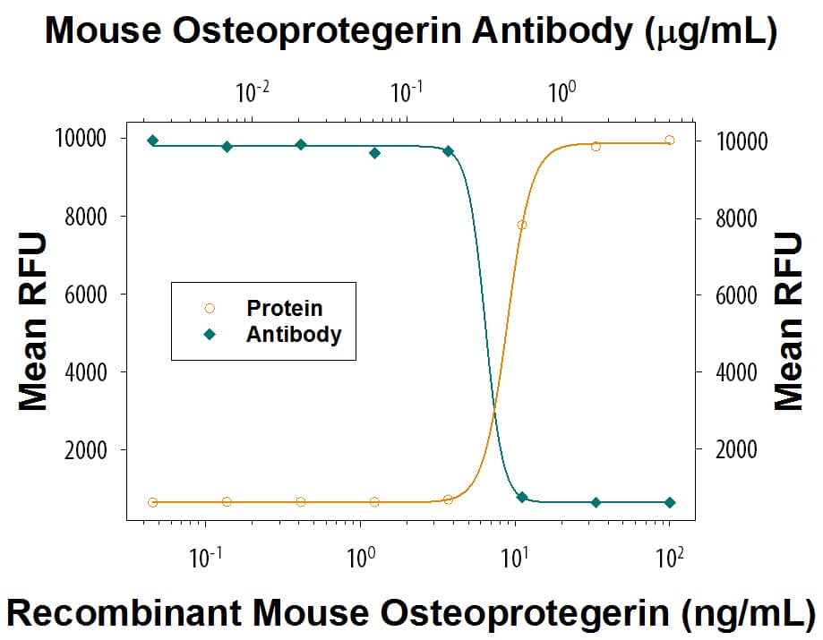
Osteoprotegerin/TNFRSF11B Inhibition of TRAIL/TNFSF10-induced Cytotoxicity and Neutralization by Mouse Osteoprotegerin/TNFRSF11B Antibody.
In the presence of the metabolic inhibitor actinomycin D (1 µg/mL), Recombinant Mouse OPG/TNFRSF11B Fc Chimera (Catalog # 459-MO) inhibits Recombinant Human TRAIL/TNFSF10 (Catalog # 375-TL) induced cytotoxicity in the L-929 mouse fibroblast cell line in a dose-dependent manner (orange line), as measured by crystal violet staining. Under these conditions, inhibition of Recombinant Human TRAIL/TNFSF10 (20 ng/mL) activity elicited by Recombinant Mouse OPG/TNFRSF11B Fc Chimera (0.1 µg/mL) is neutralized (green line) by increasing concentrations of Mouse OPG/TNFRSF11B Antigen Affinity-purified Polyclonal Antibody (Catalog # AF459). The ND50 is typically 0.15-0.9 µg/mL.Osteoprotegerin/TNFRSF11B in Mouse Stomach.
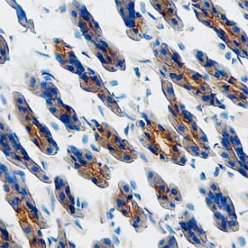
Osteoprotegerin/TNFRSF11B was detected in perfusion fixed frozen sections of mouse stomach using Goat Anti-Mouse Osteoprotegerin/TNFRSF11B Antigen Affinity-purified Polyclonal Antibody (Catalog # AF459) at 5 µg/mL overnight at 4 °C. Tissue was stained using the Anti-Goat HRP-DAB Cell & Tissue Staining Kit (brown; Catalog # CTS008) and counterstained with hematoxylin (blue). Specific staining was localized to glandular epithelial cells. View our protocol for Chromogenic IHC Staining of Frozen Tissue Sections.Detection of Mouse Osteoprotegerin/TNFRSF11B by Western Blot
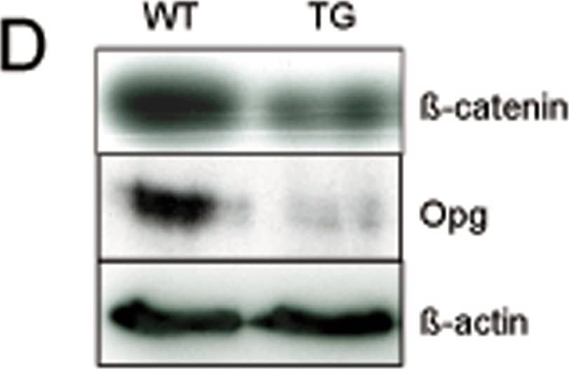
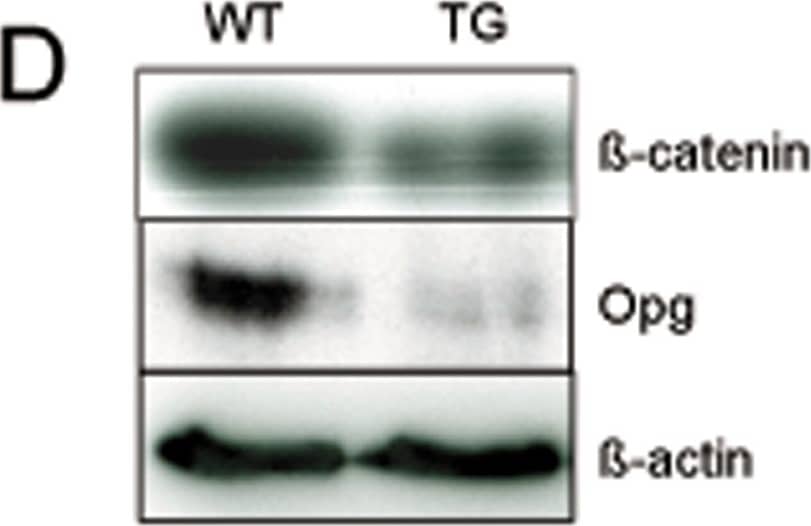
Cell-autonomous defect of Col1a1-Krm2 transgenic osteoblasts.(A) BrdU incorporation assays revealed a higher proliferation rate in primary calvarial osteoblast cultures from transgenic mice after 2 days of differentiation. (B) Von Kossa staining performed at 10 days of differentiation reveals reduced mineralization of osteoblasts from Col1a1-Krm2 mice (scale bars, 1 cm), despite higher protein content (given below). Values represent mean ± SD (n = 3). Asterisks indicate statistically significant differences. (C) Western Blot analysis of canonical Wnt signaling using primary osteoblasts from wildtype and transgenic mice following stimulation with Wnt3a for 30 minutes. (D) Western Blot analysis (left) and ELISA (right) showing decreased Opg levels in cellular extracts and conditioned medium of osteoblasts from Col1a1-Krm2 mice. (E) Quantitative RT-PCR (left) and ELISA (right) demonstrating that the reduced expression of Tnfrsf11b in bones of 6 weeks old Col1a1-Krm2 mice results in decreased Opg serum levels. All bars represent mean ± SD (n = 4). Asterisks indicate statistically significant differences. Image collected and cropped by CiteAb from the following open publication (https://pubmed.ncbi.nlm.nih.gov/20436912), licensed under a CC-BY license. Not internally tested by R&D Systems.Applications for Mouse Osteoprotegerin/TNFRSF11B Antibody
Immunohistochemistry
Sample: Perfusion fixed frozen sections of mouse stomach
Neutralization
Reviewed Applications
Read 4 reviews rated 4.5 using AF459 in the following applications:
Formulation, Preparation, and Storage
Purification
Reconstitution
Formulation
Shipping
Stability & Storage
- 12 months from date of receipt, -20 to -70 °C as supplied.
- 1 month, 2 to 8 °C under sterile conditions after reconstitution.
- 6 months, -20 to -70 °C under sterile conditions after reconstitution.
Background: Osteoprotegerin/TNFRSF11B
Osteoprotegerin (OPG)/Osteoclastogenesis Inhibitory Factor (OCIF) is member of the tumor necrosis factor receptor superfamily that lacks any apparent cell-association motifs and exists as a soluble secreted protein. In the new TNF superfamily nomenclature, OPG is referred to as TNFRSF11B. OPG was originally isolated by sequence homology as a TNF receptor family protein during a fetal rat intestine cDNA-sequencing project and subsequently shown to be involved in the regulation of bone density. OCIF was initially purified from the conditioned medium of human embryonic fibroblasts based on its ability to inhibit osteoclast development. Comparison of the amino-acid sequences of human OPG and OCIF proteins revealed their identity. The amino-terminal half of OPG contains four cysteine-rich repeats characteristic of TNF receptor family members. The carboxy-terminal of OPG/OCIF was found to contain two death domain homologous regions in tandem. Human and mouse OPG share approximately 84% and 94% amino acid sequence identity, respectively, with the rat OPG. Natural OPG/OCIF has been found to exist predominantly as disulfide-linked dimers. Two TNF superfamily ligands, including the membrane proteins OPG ligand/TRANCE (tumor necrosis factor-related activation-induced cytokine)/ODF (osteoclast differentiation factor)/RANKL (receptor activator of NF-kappaB ligand) and TRAIL (TNF-related apoptosis-inducing ligand)/APO-2 ligand, have been shown to be the cellular ligands for OPG/OCIF. Each of these ligands has been shown to interact with additional TNF receptor family members, including RANK (with TRANCE) and TRAIL receptors 1-4 (with TRAIL). The roles of these receptor-ligands in osteoclastogenesis, apoptosis and in the immune system remains to be elucidated.
References
- Lacey, D.L. et al. (1998) Cell 93:165.
- Emery, J.G. et al. (1998) J. Biol. Chem. 273:14363.
- Yasuda, H. et al. (1998) Proc. Natl. Acad. Sci. USA 95:3597.
Alternate Names
Gene Symbol
UniProt
Additional Osteoprotegerin/TNFRSF11B Products
Product Documents for Mouse Osteoprotegerin/TNFRSF11B Antibody
Product Specific Notices for Mouse Osteoprotegerin/TNFRSF11B Antibody
For research use only


