Detection of Mouse Plexin C1 by Immunocytochemistry/Immunofluorescence
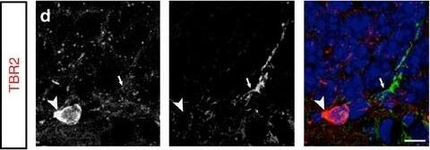
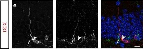
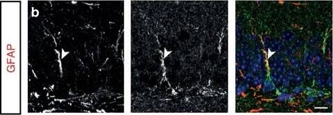
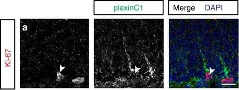
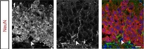
PlexinC1 expression in the subgranular zone is largely confined to early progenitors.(a–f) Double immunohistochemistry for plexinC1 and Ki-67 (a), glial fibrillary acidic protein (GFAP; b), sex determining region Y-box 2 (Sox2; c), t-box brain 2 (TBR2; d), doublecortin (DCX; e) or neuronal nuclei (NeuN; f) in sections of the adult mouse dentate gyrus (DG). The majority of plexinC1-positive cells express Ki-67, GFAP and Sox2 but not TBR2, DCX or NeuN. Arrowheads indicate cells expressing specific markers. Arrows indicate plexinC1-positive cells not expressing the indicated marker. (g,h) Quantification of the fraction of cells expressing a specific marker that also express plexinC1 (g) or the fraction of plexinC1-positive cells that also express the indicated marker protein (h). Data are presented as means±s.e.m. n≥3 (mice). (i) Schematic representation of lineage-marker and plexinC1 expression in cell lineage subtypes during neuronal differentiation in the adult DG. Ki-67 is a marker for proliferating cells during all active phases of the cell cycle. PlexinC1 is expressed in GFAP-positive and Sox2-positive radial glia-like cells (RGLs) and early intermediate progenitor cells (IPCs). Ki-67-positive proliferating cells express plexinC1. In contrast, only a small fraction of TBR2-, DCX- or NeuN-positive intermediate progenitor cells and (immature) granule cells express plexinC1. Scale bars: 10 μm. Image collected and cropped by CiteAb from the following publication (https://www.nature.com/articles/ncomms14666), licensed under a CC-BY license. Not internally tested by R&D Systems.
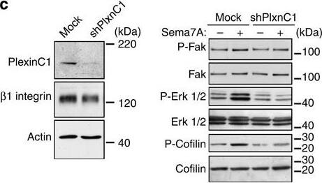
Detection of Mouse Plexin C1 by Knockdown Validated
Sema7A induces the retraction of GnRH neurites through the PlexinC1 transduction pathway and Rap1 inactivation.(a) Representative images of semaphorin-induced neurite retraction in GnV3 neurons transfected with an empty vector (mock), a construct encoding a PlexinC1 short hairpin RNA (GnV3 shRNA PlexinC1) or a Rap1-V12 plasmid encoding a constitutively active form of Rap1. One day following Sema7A treatment (250 ng ml−1), cells were labelled for F-actin (red) to visualize cytoskeletal changes. (b) Quantitative analysis of neurite length in GnV3 cells under different treatment conditions (n=5 independent experiments, n=303 mock-transfected control cells, n=303 mock-transfected cells after Sema7A treatment, unpaired Student’s t-test, ***P<0.0001; n=267 shPlxnC1-treated cells, n=235 shPlxnC1+Sema7A-treated cells, unpaired Student’s t-test, P>0.05; n=131 Rap1-V12-transfected cells, n=115 Rap1-V12-transfected cells+Sema7A, unpaired Student’s t-test, P>0.05). (c) Immunoblotting for markers indicated using transfected GnV3 cells. (d–f) Saline, Sema7A or Sema7A+PlexinC1 was infused (0.2 μg μl−1, 0.5 μl h−1 for 7 days) by stereotaxic implantation of a 28-gauge infusion cannula connected to a subcutaneously implanted mini-osmotic pump in the ME of cycling female rats. Representative oestrous cycle profiles showing the disruption of oestrous cyclicity by the infusion of Sema7A but not of PBS into the ME. Infusion was started on day 9 (downward arrow) and ended 7 days later (upward arrow), when pump contents were exhausted. Die, Diestrus; Es, estrus; Pro, proestrus. (g) Quantitative analysis of alterations in ovarian cyclicity (percentage of time in diestrus) caused by PBS, Sema7A or Sema7A+soluble PlexinC1 infusion into the rat ME (n=6 animals per group, Kruskal–Wallis test, **P<0.005). Scale bar, (a) 10 μm. Image collected and cropped by CiteAb from the following publication (https://pubmed.ncbi.nlm.nih.gov/25721933), licensed under a CC-BY license. Not internally tested by R&D Systems.
Detection of Mouse Plexin C1 by Immunocytochemistry/Immunofluorescence
PlexinC1 expression in the subgranular zone is largely confined to early progenitors.(a–f) Double immunohistochemistry for plexinC1 and Ki-67 (a), glial fibrillary acidic protein (GFAP; b), sex determining region Y-box 2 (Sox2; c), t-box brain 2 (TBR2; d), doublecortin (DCX; e) or neuronal nuclei (NeuN; f) in sections of the adult mouse dentate gyrus (DG). The majority of plexinC1-positive cells express Ki-67, GFAP and Sox2 but not TBR2, DCX or NeuN. Arrowheads indicate cells expressing specific markers. Arrows indicate plexinC1-positive cells not expressing the indicated marker. (g,h) Quantification of the fraction of cells expressing a specific marker that also express plexinC1 (g) or the fraction of plexinC1-positive cells that also express the indicated marker protein (h). Data are presented as means±s.e.m. n≥3 (mice). (i) Schematic representation of lineage-marker and plexinC1 expression in cell lineage subtypes during neuronal differentiation in the adult DG. Ki-67 is a marker for proliferating cells during all active phases of the cell cycle. PlexinC1 is expressed in GFAP-positive and Sox2-positive radial glia-like cells (RGLs) and early intermediate progenitor cells (IPCs). Ki-67-positive proliferating cells express plexinC1. In contrast, only a small fraction of TBR2-, DCX- or NeuN-positive intermediate progenitor cells and (immature) granule cells express plexinC1. Scale bars: 10 μm. Image collected and cropped by CiteAb from the following publication (https://www.nature.com/articles/ncomms14666), licensed under a CC-BY license. Not internally tested by R&D Systems.

