Mouse/Rat DLL1 Antibody
R&D Systems, part of Bio-Techne | Catalog # AF3970

Key Product Details
Species Reactivity
Validated:
Mouse, Rat
Cited:
Human, Mouse, Avian - Chicken
Applications
Validated:
CyTOF-ready, Flow Cytometry, Immunohistochemistry, Western Blot
Cited:
Immunocytochemistry, Immunohistochemistry, Immunohistochemistry-Frozen, Western Blot
Label
Unconjugated
Antibody Source
Polyclonal Sheep IgG
Product Specifications
Immunogen
Mouse myeloma cell line NS0-derived recombinant rat DLL1
Gln18-Trp537 (predicted)
Accession # P97677
Gln18-Trp537 (predicted)
Accession # P97677
Specificity
Detects rat and mouse DLL1 in direct ELISAs and Western blots. In direct ELISAs, approximately 50% cross-reactivity with recombinant human DLL‑1 is observed.
Clonality
Polyclonal
Host
Sheep
Isotype
IgG
Scientific Data Images for Mouse/Rat DLL1 Antibody
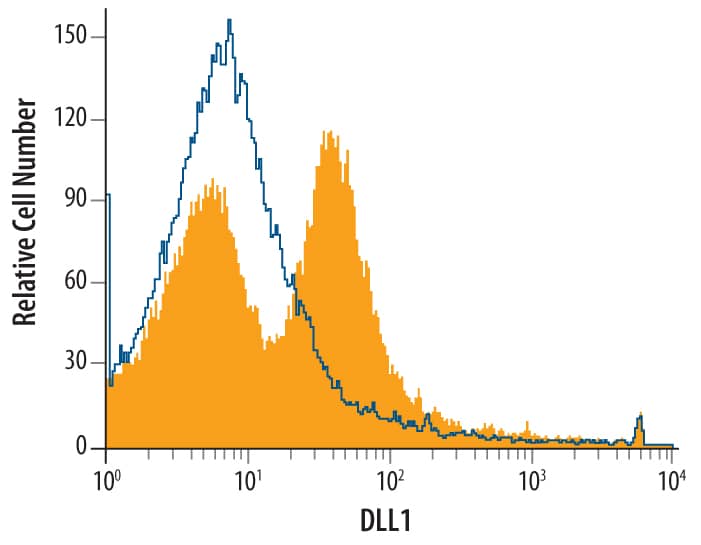
Detection of DLL1 in Mouse Splenocytes by Flow Cytometry.
Mouse splenocytes were stained with Sheep Anti-Mouse/Rat DLL1 Antigen Affinity-purified Polyclonal Antibody (Catalog # AF3970, filled histogram) or control antibody (5-001-A, open histogram), followed by NorthernLights™ 637-conjugated Anti-Sheep IgG Secondary Antibody (NL011).DLL1 in Embryonic Mouse Stomach.
DLL1 was detected in immersion fixed frozen sections of embryonic mouse stomach (E13.5) using 10 µg/mL Sheep Anti-Mouse/Rat DLL1 Antigen Affinity-purified Polyclonal Antibody (Catalog # AF3970) overnight at 4 °C. Tissue was stained with the NorthernLights™ 557-conjugated Anti-Sheep IgG Secondary Antibody (red; NL010) and counterstained with DAPI (blue). View our protocol for Fluorescent IHC Staining of Frozen Tissue Sections.Detection of Mouse DLL1 by Immunocytochemistry/Immunofluorescence
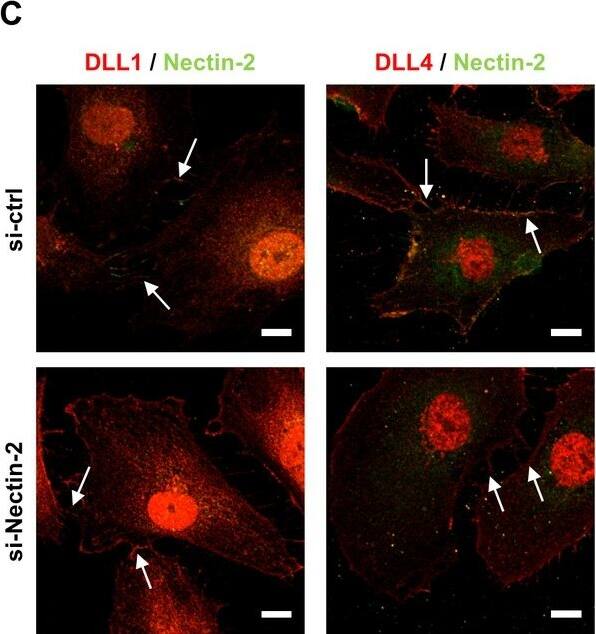
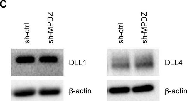
Mpdz does not affect cell cell junction assembly.(A, B) HUVECs were either transduced with lentivirus expressing GFP (sh-ctrl) or with lentivirus expressing shRNA against MPDZ (sh-MPDZ). Cells were cultured under sparse conditions (A) or confluent conditions (B). After PFA fixation cells were stained for DLL1 and Nectin-2 or DLL4 and Nectin-2 and counterstained with DAPI. Images were acquired with the confocal microscope LSM 700. Arrow indicates co-localization of DLL1/4 with Nectin-2 at the cell membrane. Arrow head indicates diminished co-localization at the cell membrane. Scale bar: 10 µm. (C) HUVECs were either transfected with control siRNA (si-ctrl) or with siRNA against Nectin-2 (si-Nectin-2). After PFA fixation cells were stained for DLL1 and Nectin-2 or DLL4 and Nectin-2. Images were acquired with the confocal microscope LSM 700. Arrow indicates localization of DLL1/4 at the cell membrane.Scale bar: 10 µm. Image collected and cropped by CiteAb from the following publication (https://pubmed.ncbi.nlm.nih.gov/29620522), licensed under a CC-BY license. Not internally tested by R&D Systems.Applications for Mouse/Rat DLL1 Antibody
Application
Recommended Usage
CyTOF-ready
Ready to be labeled using established conjugation methods. No BSA or other carrier proteins that could interfere with conjugation.
Flow Cytometry
1 µg/106 cells
Sample: CHO cells transfected with DLL1
Sample: CHO cells transfected with DLL1
Immunohistochemistry
0.5-15 µg/mL
Sample: Immersion fixed frozen sections of embryonic mouse stomach (E13.5)
Sample: Immersion fixed frozen sections of embryonic mouse stomach (E13.5)
Western Blot
0.1 µg/mL
Sample: Recombinant Rat DLL1 Fc Chimera (Catalog # 3970-DL)
Sample: Recombinant Rat DLL1 Fc Chimera (Catalog # 3970-DL)
Formulation, Preparation, and Storage
Purification
Antigen Affinity-purified
Reconstitution
Reconstitute at 0.2 mg/mL in sterile PBS. For liquid material, refer to CoA for concentration.
Formulation
Lyophilized from a 0.2 μm filtered solution in PBS with Trehalose. See Certificate of Analysis for details.
*Small pack size (-SP) is supplied either lyophilized or as a 0.2 µm filtered solution in PBS.
*Small pack size (-SP) is supplied either lyophilized or as a 0.2 µm filtered solution in PBS.
Shipping
Lyophilized product is shipped at ambient temperature. Liquid small pack size (-SP) is shipped with polar packs. Upon receipt, store immediately at the temperature recommended below.
Stability & Storage
Use a manual defrost freezer and avoid repeated freeze-thaw cycles.
- 12 months from date of receipt, -20 to -70 °C as supplied.
- 1 month, 2 to 8 °C under sterile conditions after reconstitution.
- 6 months, -20 to -70 °C under sterile conditions after reconstitution.
Background: DLL1
gamma-secretase, enabling the cytoplasmic domain to migrate to the nucleus (5). DLL1 localizes to adherens junctions on neuronal processes through its association with the scaffolding protein MAGI1 (6). DLL1 is widely expressed, and it plays an important role in embryonic somite formation, cochlear hair cell differentiation, lymphocyte differentiation, and the maintenance of neural and myogenic progenitor cells (4, 7-13). The upregulation of DLL1 in arterial endothelial cells following injury or angiogenic stimulation is central to postnatal arteriogenesis (14). DLL1 is also overexpressed in cervical carcinoma and glioma and contributes to tumor progression (15-16).
References
- Bettenhausen, B. et al. (1995) Development 121:2407.
- Dyczynska, E. et al. (2007) J. Biol. Chem. 282:436.
- Karanu, F.N. et al. (2001) Blood 97:1960.
- Sun, D. et al. (2008) J. Cell Sci. 121:3815.
- Ikeuchi, T. and S.S. Sisodia (2003) J. Biol. Chem. 278:7751.
- Mizuhara, E. et al. (2005) J. Biol. Chem. 280:26499.
- Takahashi, Y. et al. (2003) Development 130:4259.
- Teppner, I. et al. (2007) BMC Dev. Biol. 7:68.
- Kiernan, A.E. et al. (2005) Development 132:4353.
- Schmitt, T.M. and J.C. Zuniga-Pflucker (2002) Immunity 17:749.
- Hozumi, K. et al. (2004) Nat. Immunol. 5:638.
- Shimojo, H. et al. (2008) Neuron 58:52.
- Schuster-Gossler, K. et al. (2007) Proc. Natl. Acad. Sci. USA 104:537.
- Limbourg, A. et al. (2007) Circ. Res. 100:363.
- Purow, B.W. et al. (2005) Cancer Res. 65:2353.
- Gray, G.E. et al. (1999) Am. J. Pathol. 154:785.
Long Name
Delta-like 1
Alternate Names
Delta 1
Gene Symbol
DLL1
UniProt
Additional DLL1 Products
Product Documents for Mouse/Rat DLL1 Antibody
Product Specific Notices for Mouse/Rat DLL1 Antibody
For research use only
Loading...
Loading...
Loading...
Loading...
Loading...


