Mouse/Rat M-Cadherin/Cadherin-15 Antibody
R&D Systems, part of Bio-Techne | Catalog # AF7677

Key Product Details
Species Reactivity
Validated:
Cited:
Applications
Validated:
Cited:
Label
Antibody Source
Product Specifications
Immunogen
Val22-Ala605 (Arg159His)
Accession # P33146
Specificity
Clonality
Host
Isotype
Scientific Data Images for Mouse/Rat M-Cadherin/Cadherin-15 Antibody
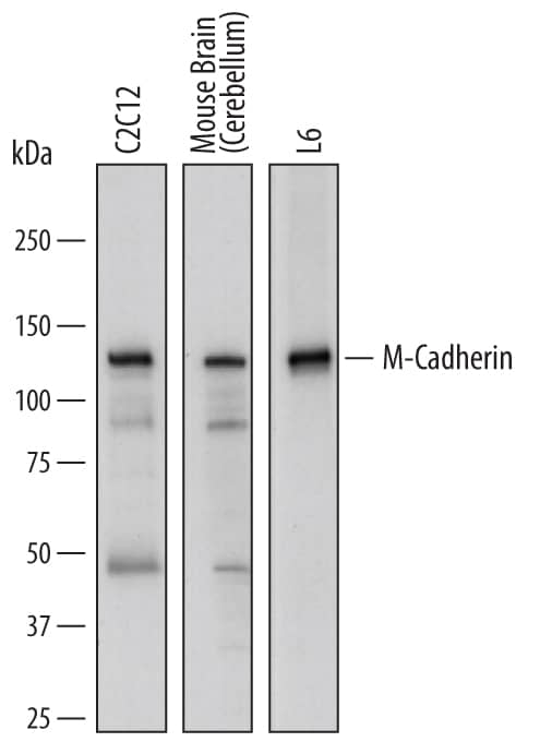
Detection of Mouse and Rat M‑Cadherin/ Cadherin‑15 by Western Blot.
Western blot shows lysates of C2C12 mouse myoblast cell line, mouse brain (cerebellum) tissue, and L6 rat myoblast cell line. PVDF membrane was probed with 1 µg/mL of Sheep Anti-Mouse/Rat M-Cadherin/Cadherin-15 Antigen Affinity-purified Polyclonal Antibody (Catalog # AF7677) followed by HRP-conjugated Anti-Sheep IgG Secondary Antibody (Catalog # HAF016). A specific band was detected for M-Cadherin/Cadherin-15 at approximately 125 kDa (as indicated). This experiment was conducted under reducing conditions and using Immunoblot Buffer Group 1.M‑Cadherin/Cadherin‑15 in Human Embryonic Muscle.
M-Cadherin/Cadherin-15 was detected in perfusion fixed frozen sections of human embryonic muscle using Sheep Anti-Mouse/Rat M-Cadherin/Cadherin-15 Antigen Affinity-purified Polyclonal Antibody (Catalog # AF7677) at 1.7 µg/mL overnight at 4 °C. Tissue was stained using the NorthernLights™ 557-conjugated Anti-Sheep IgG Secondary Antibody (red; Catalog # NL010) and counterstained with DAPI (blue). Specific staining was localized to plasma membrane. View our protocol for Fluorescent IHC Staining of Frozen Tissue Sections.Applications for Mouse/Rat M-Cadherin/Cadherin-15 Antibody
Immunohistochemistry
Sample: Perfusion fixed frozen sections of human embryonic muscle
Western Blot
Sample: C2C12 mouse myoblast cell line, mouse brain (cerebellum) tissue, and L6 rat myoblast cell line
Formulation, Preparation, and Storage
Purification
Reconstitution
Formulation
Shipping
Stability & Storage
- 12 months from date of receipt, -20 to -70 °C as supplied.
- 1 month, 2 to 8 °C under sterile conditions after reconstitution.
- 6 months, -20 to -70 °C under sterile conditions after reconstitution.
Background: M-Cadherin/Cadherin-15
CDH-15 (Cadherin 15; also M-cadherin, Muscle cadherin and Cadherin-14) is a 125-127 kDa atypical member of the classical cadherin family, cadherin superfamily of molecules. It is expressed by muscle satellite cells, cells of the embryonic myotome, and hematopoietic bone marrow stem cells. CDH-15 appears to bind homotypically in trans, thus allowing for the identification and subsequent fusion of myoblast precursors, particularly those in slow-twitch (or red fiber) muscles. This is accompanied by a downregulation of mitochondrial induced apoptosis. Mouse CDH-15 is synthesized as a 784 amino acid (aa) preproprecursor. It contains a 21 aa signal sequence, a 38 aa propeptide, and a 725 aa mature region. The mature region is expressed as a type I transmembrane glycoprotein that possesses a 546 aa extracellular region (aa 60-605) and a 159 aa cytoplasmic domain (aa 626-784). The extracellular region shows five consecutive cadherin domains. Over aa 22-605, mouse CDH-15 shares 88% and 97% aa sequence identity with human and rat CDH-15, respectively.
Long Name
Alternate Names
Gene Symbol
UniProt
Additional M-Cadherin/Cadherin-15 Products
Product Documents for Mouse/Rat M-Cadherin/Cadherin-15 Antibody
Product Specific Notices for Mouse/Rat M-Cadherin/Cadherin-15 Antibody
For research use only

