Mouse/Rat MMP-2 Antibody
R&D Systems, part of Bio-Techne | Catalog # AF1488


Key Product Details
Validated by
Species Reactivity
Validated:
Cited:
Applications
Validated:
Cited:
Label
Antibody Source
Product Specifications
Immunogen
Ile34-Cys662
Accession # P33434
Specificity
Clonality
Host
Isotype
Scientific Data Images for Mouse/Rat MMP-2 Antibody
MMP‑2 in Mouse Thymus.
MMP-2 was detected in perfusion fixed frozen sections of mouse thymus using Goat Anti-Mouse/ Rat MMP-2 Antigen Affinity-purified Polyclonal Antibody (Catalog # AF1488) at 15 µg/mL overnight at 4 °C. Tissue was stained using the Anti-Goat HRP-DAB Cell & Tissue Staining Kit (brown; Catalog # CTS008) and counterstained with hematoxylin (blue). View our protocol for Chromogenic IHC Staining of Frozen Tissue Sections.Detection of Mouse MMP-2 by Immunohistochemistry-Paraffin
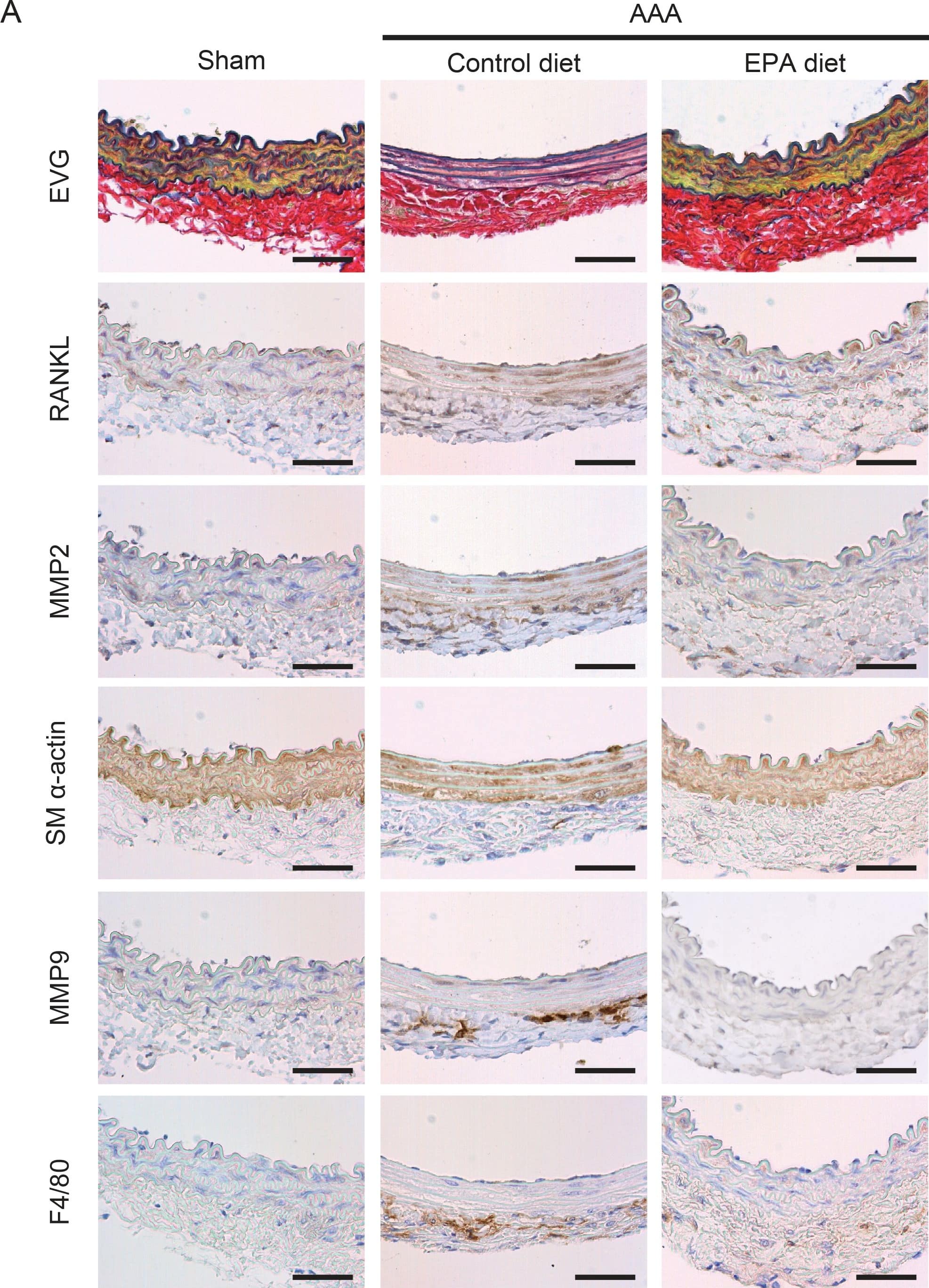
MMP2, MMP9, and RANKL expression in AAAs.A. Immunohistochemical staining for indicated proteins of serial sections of aortas one week after CaCl2 treatment. Elastic van Gieson staining is also shown. SM alpha-actin and F4/80 were stained to locate SMCs and macrophages, respectively. Shown are representative images of 4 or more samples in each group. Scale bars, 50 µm. B. Relative positive staining area of MMP2, MMP9, and RANKL in sections from control diet and EPA diet groups. n = 4–5. *P<0.05. Image collected and cropped by CiteAb from the following open publication (https://dx.plos.org/10.1371/journal.pone.0096286), licensed under a CC-BY license. Not internally tested by R&D Systems.Applications for Mouse/Rat MMP-2 Antibody
Immunohistochemistry
Sample: Perfusion fixed frozen sections of mouse thymus
Immunoprecipitation
Sample: Conditioned cell culture medium spiked with Recombinant Mouse/Rat MMP‑2 (Catalog # 924‑MP), see our available Western blot detection antibodies
Western Blot
Sample: Recombinant Mouse/Rat MMP-2 (Catalog # 924-MP)
Formulation, Preparation, and Storage
Purification
Reconstitution
Formulation
Shipping
Stability & Storage
- 12 months from date of receipt, -20 to -70 °C as supplied.
- 1 month, 2 to 8 °C under sterile conditions after reconstitution.
- 6 months, -20 to -70 °C under sterile conditions after reconstitution.
Background: MMP-2
Matrix metalloproteinases are a family of zinc and calcium dependent endopeptidases with the combined ability to degrade all the components of the extracellular matrix. MMP-2 (gelatinase A), a type IV collagenase, can degrade a broad range of substrates including type IV, V, VII and X collagens as well as elastin and fibronectin. It is believed to act synergistically with interstitial collagenase (MMP-1) in the degradation of fibrillar collagens as it degrades their denatured gelatin forms. MMP-2 has been shown to be associated with many connective tissue cells as well as neutrophils, macrophages and monocytes. Structurally, MMP-2 may be divided into several distinct domains: a pro-domain which is cleaved upon activation; a catalytic domain containing the zinc binding site; a fibronectin-like domain thought to play a role in substrate targeting; and a carboxyl terminal (hemopexin-like) domain containing 2 N-linked glycosylation sites. The amino acid sequences of the proenzymes are identical between mouse and rat.
Long Name
Alternate Names
Gene Symbol
UniProt
Additional MMP-2 Products
Product Documents for Mouse/Rat MMP-2 Antibody
Product Specific Notices for Mouse/Rat MMP-2 Antibody
For research use only
