Rat Neuropilin-1 Alexa Fluor® 700-conjugated Antibody
R&D Systems, part of Bio-Techne | Catalog # FAB566N


Key Product Details
Species Reactivity
Validated:
Cited:
Applications
Validated:
Cited:
Label
Antibody Source
Product Specifications
Immunogen
Phe22-Ala810 (Lys811Arg), Ser829-Asp854
Accession # Q9QWJ9
Specificity
Clonality
Host
Isotype
Scientific Data Images for Rat Neuropilin-1 Alexa Fluor® 700-conjugated Antibody
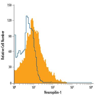
Detection of Neuropilin‑1 in bEnd.3 Mouse Cell Line by Flow Cytometry.
bEnd.3 mouse endothelioma cell line was stained with Goat Anti-Mouse/Rat Neuropilin-1 Alexa Fluor® 700-conjugated Antigen Affinity-purified Polyclonal Antibody (Catalog # FAB566N, filled histogram) or isotype control antibody (Catalog # IC108N, open histogram). View our protocol for Staining Membrane-associated Proteins.Detection of Neuropilin‑1 in PC‑12 Rat Cell Line by Flow Cytometry.
PC-12 rat adrenal pheochromocytoma cell line was stained with Goat Anti-Mouse/Rat Neuropilin-1 Alexa Fluor® 700-conjugated Antigen Affinity-purified Polyclonal Antibody (Catalog # FAB566N, filled histogram) or isotype control antibody (Catalog # IC108N, open histogram). View our protocol for Staining Membrane-associated Proteins.Detection of Mouse Neuropilin-1 by Flow Cytometry
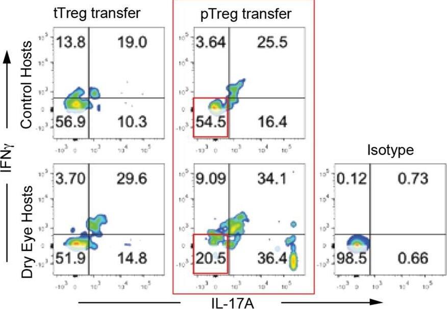
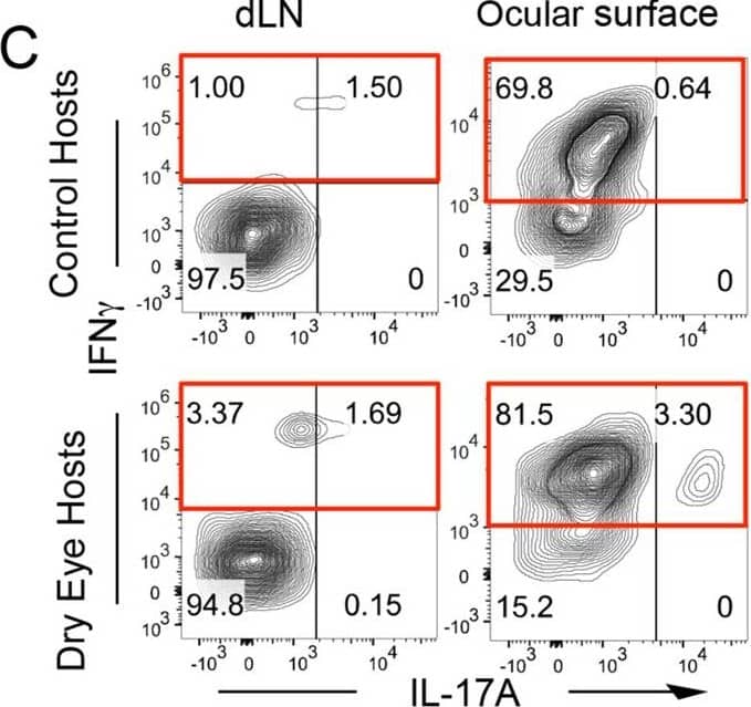
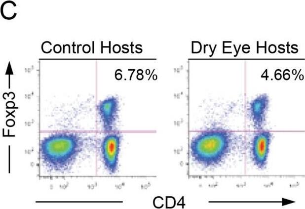
pTregs show a higher susceptibility to convert into exFoxp3 cells after transfer into dry eye hosts. pTregs (CD4+CD25+Nrp-1−) and tTregs (CD4+CD25+Nrp-1+) from transgenic dry eye disease (DED) mice (Foxp3-GFP×R26-RFP) were adoptively transferred into control and DED transplant recipients. (A and B) Draining lymph nodes were harvested at day 14 post-transplantation. Single cell suspensions were analyzed by flow cytometry. Frequencies of exFoxp3 cells (GFP−RFP+) from control and DED hosts that received (A) pTregs (59.02 ± 0.92% vs. 78.26 ± 0.69%, n = 4, ***p < 0.0001 unpaired two-tailed Student’s t test) or (B) tTregs was assessed. (C) Representative flow cytometry plots showing frequencies of IL-17 and IFN gamma-expressing exFoxp3 cells in draining lymph nodes from control and dry eye hosts. (D) Nrp1+ tTregs or Nrp1− pTregs from hosts with accepted allografts were adoptively transferred to dry eye hosts, and graft survival was assessed for 8 weeks (n = 10, χ2 = 0.6087, p = 0.4353). Image collected and cropped by CiteAb from the following publication (https://pubmed.ncbi.nlm.nih.gov/29728574), licensed under a CC-BY license. Not internally tested by R&D Systems.Applications for Rat Neuropilin-1 Alexa Fluor® 700-conjugated Antibody
Blockade of Receptor-ligand Interaction
CyTOF-ready
Flow Cytometry
Sample: bEnd.3 mouse endothelioma cell line and PC‑12 rat adrenal pheochromocytoma cell line
Formulation, Preparation, and Storage
Purification
Formulation
Shipping
Stability & Storage
- 12 months from date of receipt, 2 to 8 °C as supplied.
Background: Neuropilin-1
Alternate Names
Gene Symbol
UniProt
Additional Neuropilin-1 Products
Product Specific Notices for Rat Neuropilin-1 Alexa Fluor® 700-conjugated Antibody
* This product or the use of this product is covered by U.S. Patents owned by The Regents of the University of California. This product is for research use only and is not to be used for commercial purposes. Use of this product to produce products for sale or for diagnostic, therapeutic or drug discovery purposes is prohibited. In order to obtain a license to use this product for such purposes, contact The Regents of the University of California.
U.S. Patent # 6,054,293, 6,623,738, and other U.S. and international patents pending.
This product is provided under an agreement between Life Technologies Corporation and R&D Systems, Inc, and the manufacture, use, sale or import of this product is subject to one or more US patents and corresponding non-US equivalents, owned by Life Technologies Corporation and its affiliates. The purchase of this product conveys to the buyer the non-transferable right to use the purchased amount of the product and components of the product only in research conducted by the buyer (whether the buyer is an academic or for-profit entity). The sale of this product is expressly conditioned on the buyer not using the product or its components (1) in manufacturing; (2) to provide a service, information, or data to an unaffiliated third party for payment; (3) for therapeutic, diagnostic or prophylactic purposes; (4) to resell, sell, or otherwise transfer this product or its components to any third party, or for any other commercial purpose. Life Technologies Corporation will not assert a claim against the buyer of the infringement of the above patents based on the manufacture, use or sale of a commercial product developed in research by the buyer in which this product or its components was employed, provided that neither this product nor any of its components was used in the manufacture of such product. For information on purchasing a license to this product for purposes other than research, contact Life Technologies Corporation, Cell Analysis Business Unit, Business Development, 29851 Willow Creek Road, Eugene, OR 97402, Tel: (541) 465-8300. Fax: (541) 335-0354.
For research use only


