Mouse/Rat Notch-1 Antibody
R&D Systems, part of Bio-Techne | Catalog # AF1057


Key Product Details
Species Reactivity
Validated:
Cited:
Applications
Validated:
Cited:
Label
Antibody Source
Product Specifications
Immunogen
Arg20-Glu488 (Ala208Thr, Asp334Glu)
Accession # Q07008
Specificity
Clonality
Host
Isotype
Endotoxin Level
Scientific Data Images for Mouse/Rat Notch-1 Antibody
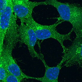
Notch‑1 in Rat Cortical Stem Cells.
Notch-1 was detected in immersion fixed undifferentiated rat cortical stem cells using Goat Anti-Mouse/Rat Notch-1 Antigen Affinity-purified Polyclonal Antibody (Catalog # AF1057) at 10 µg/mL for 3 hours at room temperature. Cells were stained using the NorthernLights™ 493-conjugated Anti-Goat IgG Secondary Antibody (green; Catalog # NL003) and counterstained with DAPI (blue). Specific staining was localized to cell surfaces. View our protocol for Fluorescent ICC Staining of Stem Cells on Coverslips.Notch‑1 in Mouse Cortical Stem Cells.
Notch-1 was detected in immersion fixed undifferentiated mouse cortical stem cells using Goat Anti-Mouse/Rat Notch-1 Antigen Affinity-purified Polyclonal Antibody (Catalog # AF1057) at 10 µg/mL for 3 hours at room temperature. Cells were stained using the NorthernLights™ 493-conjugated Anti-Goat IgG Secondary Antibody (green; Catalog # NL003) and counterstained with DAPI (blue). Specific staining was localized to cell surfaces. View our protocol for Fluorescent ICC Staining of Stem Cells on Coverslips.Notch-1 in C2C12 Mouse Cell Line.
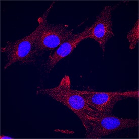
Notch-1 was detected in immersion fixed C2C12 mouse myoblast cell line using Goat Anti-Mouse/Rat Notch-1 Antigen Affinity-purified Polyclonal Antibody (Catalog # AF1057) at 15 µg/mL for 3 hours at room temperature. Cells were stained using the NorthernLights™ 557-conjugated Anti-Goat IgG Secondary Antibody (red; NL001) and counterstained with DAPI (blue). Specific staining was localized to cytoplasm. Staining was performed using our protocol for Fluorescent ICC Staining of Non-adherent Cells.Applications for Mouse/Rat Notch-1 Antibody
Blockade of Receptor-ligand Interaction
CyTOF-ready
Flow Cytometry
Sample: Rat cortical stem cells
Immunocytochemistry
Sample: Immersion fixed undifferentiated rat and mouse cortical stem cells and C2C12 mouse myoblast cell line
Immunohistochemistry
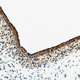
Sample: Immersion fixed paraffin-embedded sections of rat embryo (13 d.p.c.)
Western Blot
Sample: Recombinant Rat Notch-1 Fc Chimera (Catalog # 1057-TK)
Formulation, Preparation, and Storage
Purification
Reconstitution
Formulation
Shipping
Stability & Storage
- 12 months from date of receipt, -20 to -70 °C as supplied.
- 1 month, 2 to 8 °C under sterile conditions after reconstitution.
- 6 months, -20 to -70 °C under sterile conditions after reconstitution.
Background: Notch-1
Rat Notch-1 is a 300 kDa, type I transmembrane glycoprotein involved in a number of early-event developmental processes (1). In both vertebrates and invertebrates, Notch signaling is important for specifying cell fates and for defining boundaries between different cell types. The molecule is synthesized as a 2531 amino acid (aa) precursor that contains an 18 aa signal sequence, a 1705 aa extracellular region, a 23 aa transmembrane (TM) segment and a 785 aa cytoplasmic domain (2). The large Notch-1 extracellular domain has 36 EGF‑like repeats followed by three notch/Lin-12 repeats. Of the 36 EGF‑like repeats, the 11th and 12th EGF‑like repeats have been shown to be both necessary and sufficient for binding the ligands Delta and Serrate, in Drosophila (3). The Notch-1 cytoplasmic domain contains six ankyrin repeats, a glutamine-rich domain and a PEST sequence. The Notch-1 receptor undergoes post-translational proteolytic cleavage by a furin-like enzyme to form a heterodimer of the 1635 aa ligand binding extracellular region and the 877 aa transmembrane protein (4). Upon ligand binding, additional sequential proteolysis by TNF-converting enzyme and the Presenilin-dependent gamma-secretase results in the release of the Notch intracellular domain (NCID) which translocates into the nucleus where it functions as a transcription activator to initiate transcription of Notch-responsive genes (5). An alternative Notch signaling pathway that is mediated by the full-length form of Notch that has not been cleaved by the furin-like enzyme has also been reported (6). The rat Notch-1 extracellular domain shows 86% and 97% aa identity to human and mouse Notch-1 extracellular domains respectively. It also exhibits 56% and 50% aa identity with rat Notch-2 and Notch-3 extracellular domains, respectively.
References
- Weinmaster, G. (2000) Curr. Opin. Genet. Dev. 10:363.
- Weinmaster, G. et al. (1991) Development 113:199.
- Rebay, I. et al. (1991) Cell 67:687.
- Rogeat, F. et al. (1998) Proc. Natl. Acad. Sci. USA 95:8108.
- Mumm, J.S. and R. Kopan (2000) Dev. Biol. 228:151.
- Bush, G. et al. (2001) Dev. Biol. 229:494.
Alternate Names
Gene Symbol
UniProt
Additional Notch-1 Products
Product Documents for Mouse/Rat Notch-1 Antibody
Product Specific Notices for Mouse/Rat Notch-1 Antibody
For research use only


