Mouse/Rat SP-D Antibody
R&D Systems, part of Bio-Techne | Catalog # AF6839

Key Product Details
Species Reactivity
Applications
Label
Antibody Source
Product Specifications
Immunogen
Ala20-Phe374
Accession # P50404
Specificity
Clonality
Host
Isotype
Scientific Data Images for Mouse/Rat SP-D Antibody
Detection of Mouse and Rat SP‑D by Western Blot.
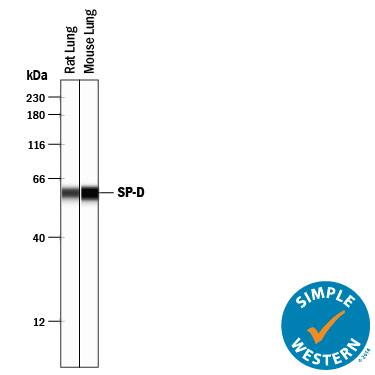
Western blot shows lysates of mouse lung tissue and rat lung tissue. PVDF membrane was probed with 2 µg/mL of Sheep Anti-Mouse/Rat SP-D Antigen Affinity-purified Polyclonal Antibody (Catalog # AF6839) followed by HRP-conjugated Anti-Sheep IgG Secondary Antibody (Catalog # HAF016). A specific band was detected for SP-D at approximately 43 kDa (as indicated). This experiment was conducted under reducing conditions and using Immunoblot Buffer Group 1.Detection of Rat and Mouse SP‑D by Simple WesternTM.
Simple Western lane view shows lysates of rat lung tissue and mouse lung tissue, loaded at 0.2 mg/mL. A specific band was detected for SP-D at approximately 60 kDa (as indicated) using 20 µg/mL of Sheep Anti-Mouse/Rat SP-D Antigen Affinity-purified Polyclonal Antibody (Catalog # AF6839) followed by 1:50 dilution of HRP-conjugated Anti-Sheep IgG Secondary Antibody (Catalog # HAF016). This experiment was conducted under reducing conditions and using the 12-230 kDa separation system.Applications for Mouse/Rat SP-D Antibody
Simple Western
Sample: Rat lung tissue and mouse lung tissue
Western Blot
Sample: Mouse lung tissue and rat lung tissue
Formulation, Preparation, and Storage
Purification
Reconstitution
Formulation
Shipping
Stability & Storage
- 12 months from date of receipt, -20 to -70 °C as supplied.
- 1 month, 2 to 8 °C under sterile conditions after reconstitution.
- 6 months, -20 to -70 °C under sterile conditions after reconstitution.
Background: SP-D
SP‑D (surfactant protein‑D; also PSP‑D) is a 43 kDa member of the collectin family of innate immune modulators (1 ‑ 5). It is constitutively secreted by alveolar lining cells and epithelium associated with tubular structures. SP‑D is found in serum, plasma, broncho‑alveolar lavage (BAL) fluid, and amniotic fluid (1, 2, 6). Lung injuries often increase release of SP‑D to the circulation (3, 6). Mouse SP‑D is synthesized as a 374 amino acid (aa) precursor. Mouse SP‑D cDNA encodes a 19 aa signal sequence and a 355 aa mature region with a 25 aa N‑terminal linking‑region, a 177 aa hydroxyproline and hydroxylysine collagen‑like domain, a 46 aa coiled‑coil segment, and a 106 aa, C‑terminal collectin‑like C‑type lectin domain (CRD) (5). Mature mouse SP‑D shares 72 ‑ 76% aa sequence identity with human, porcine, equine, canine and bovine SP‑D, and 92% with rat SP‑D. SP‑D is usually found as a glycosylated, disulfide‑linked 150 kDa alpha‑helical coiled‑coil trimer with a “head” of three symmetrical CRDs (2‑4, 7). Each CRD recognizes the hydroxides of one monosaccharide, and trimerization allows for the discrimination of monosaccharide patterns specific to microbial pathogens (4, 7, 8). Typically, SP‑D forms a higher‑order 620 kDa, X‑shaped dodecamer through N‑terminal disulfide bonds, allowing for even finer discrimination of self vs. nonself carbohydrate patterns and facilitating binding to complex antigens (1). SP‑D also binds SIRP alpha and the calreticulin/CD91 complex on macrophages (9, 10). When the ratio of antigen/pathogen to available CRDs is low, antigen can be bound without occupying all available CRDs. The free CRDs will bind to SIRP alpha, generating a signal that downmodulates the inflammatory response. During high CRD ligand binding (low SIRP alpha binding), the dodecamer rearranges to expose N‑termini that bind the calreticulin/CD91 complex, an event that initiates inflammation (1). Also, direct and indirect binding of neutrophil defensins and macrophage CD14 and TLRs to SP‑D can modulate response to viruses and bacterial lipopolysaccharides (1‑3, 11‑15). Thus, SP‑D allows for a graded response to environmental challenge and clearance of small antigenic insults without the need for a damaging inflammatory response (1‑3).
References
- Forbes, L.R. and A. Haczku (2010) Clin. Exp. Allergy 40:547.
- Kishore, U. et. al. (2006) Mol. Immunol. 43:1293.
- Hartl, D. and M. Griese (2006) Eur. J. Clin. Invest. 36:423.
- Sim, R.B. et. al. (2006) Novartis Found Symp. 279:170.
- Motwani, M. et al. (1995) J. Immunol. 155:5671.
- Honda, Y. et al. (1995) Am. J. Respir. Crit. Care Med. 152:1860.
- Hakansson, K. et. al. (1999) Structure 7:225.
- Crouch, E.C. et. al. (2006) Am. J. Respir. Cell Mol. Biol. 35:84.
- Janssen, W.J. et al. (2008) Am. J. Respir. Crit. Care Med. 178:158.
- Gardai, S.J. et al. (2003) Cell 115:13.Ohya, M. et. al. (2006) Biochemistry 45:8657.
- Ohya, M. et. al. (2006) Biochemistry 45:8657.
- Pastva, A.M. et al. (2007) Proc. Am. Thorac. Soc. 4:252.
- Sano, H. and Y. Kuroki (2005) Mol. Immunol. 42:279.
- Hartshorn, K.L. et al. (2006) J. Immunol. 176:6962.
- Yamazoe, M. et al. (2008) J. Biol. Chem. 283:35878.
Long Name
Alternate Names
Gene Symbol
UniProt
Additional SP-D Products
Product Documents for Mouse/Rat SP-D Antibody
Product Specific Notices for Mouse/Rat SP-D Antibody
For research use only

