Mouse Semaphorin 3A Antibody
R&D Systems, part of Bio-Techne | Catalog # MAB109031


Key Product Details
Species Reactivity
Applications
Label
Antibody Source
Product Specifications
Immunogen
Asn21-Lys747
Accession # O08665
Specificity
Clonality
Host
Isotype
Scientific Data Images for Mouse Semaphorin 3A Antibody
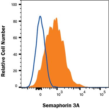
Detection of Semaphorin 3A in Mouse bEnd.3 Cell Line by Flow Cytometry.
Mouse bEnd.3 brain endothelial cell line was stained with Rat Anti-Mouse Semaphorin 3A Monoclonal Antibody (Catalog # MAB109031, filled histogram) or isotype control antibody (MAB005, open histogram), followed by Phycoerythrin-conjugated Anti-Rat IgG F(ab')2Secondary Antibody (F0105B). To facilitate intracellular staining, cells were fixed with Flow Cytometry Fixation Buffer (FC004) and permeabilized with Flow Cytometry Permeabilization/Wash Buffer (FC005). Staining was performed using our Staining Intracellular Proteins protocol.Applications for Mouse Semaphorin 3A Antibody
Intracellular Staining by Flow Cytometry
Sample: bEnd.3 mouse brain endothelial cell line
Formulation, Preparation, and Storage
Purification
Reconstitution
Formulation
Shipping
Stability & Storage
- 12 months from date of receipt, -20 to -70 °C as supplied.
- 1 month, 2 to 8 °C under sterile conditions after reconstitution.
- 6 months, -20 to -70 °C under sterile conditions after reconstitution.
Background: Semaphorin 3A
Semaphorin 3A (Sema3A; previously sem D, sema III or collapsin) is one of six Class 3 secreted semaphorins which share ~40-50% amino acid (aa) identity
(1-3). Class 3 semaphorins are potent chemorepellents that function in axon and/or vascular guidance during development (2, 3). The 772 aa mouse Sema3C contains a 20 aa signal sequence, an ~500 aa N-terminal Sema domain that forms a beta-propeller structure similar to that found in integrin molecules, a PSI domain, a furin-type cleavage site, an Ig-like domain, and a C-terminal basic domain (3, 4). Covalent dimerization plus cleavage at the C-terminus are required for activity of class 3 semaphorins (5, 6). The 95 kDa mature mouse Sema3A shares at least 95% aa identity with human, rat, equine and canine Sema3A, and 90% and 86% aa identity with chick and zebrafish Sema3A, respectively. Type 3 semaphorins transduce signals through transmembrane plexins, either directly or by binding associated neuropilin receptors (3). Sema3A signaling is transduced by plexin A1-4, indirectly via neuropilin-1 (3). Sema3A activity is mediated by small GTPases that influence actin rearrangement and integrin activity (7-9). It is important in developmental organization of central and peripheral nerves, including those in heart, lung, kidneys, bones, teeth, and visual and olfactory systems (1, 2, 10, 11). Gradients of Sema3A repel axons, but attract dendrites (11, 12). Sema3A affect vasculogenesis by inhibiting integrin function and, with Sema3F, promoting apoptosis of endothelial cells (3, 9, 12). It is thought to suppress cancer-related angiogenesis (3). In the immune system, Sema3A influences T cell proliferation, migration, response to activation, and interactions with dendritic cells (7, 13). It negatively regulates platelet activation (14). Expression of Sema3A in relevant parts of the nervous system may be increased in Alzheimer’s disease, multiple sclerosis, ischemia and schizophrenia (2).
References
- Puschel, A.W. et al. (1995) Neuron 14:941.
- Roth, L. et al. (2009) Cell. Mol. Life Sci. 66:649.
- Neufeld, G and O. Kessler (2008) Nat. Rev. Cancer 8:632.
- Gherardi, E. et al. (2004) Curr. Opin. Struct. Biol. 14:669.
- Adams, R. H. et al. (1997) EMBO J. 16:6077.
- Klosterman, A. et al. (1998) J. Biol. Sci. 273:7326.
- Lepelletier, Y. et al. (2006) Eur. J. Immunol. 36:1782.
- Schlomann, U. et al. (2009) J. Cell Sci. 122:2034.
- Serini, G. et al. (2003) Nature 424:391.
- Ieda, M. et al. (2007) Nat. Med. 13:604.
- Chen, G. et al. (2008) Nat. Neurosci. 11:36.
- Guttmann-Raviv, N. et al. (2007) J. Biol Chem. 282:26294.
- Lepelletier, Y. et al. (2007) Proc. Natl. Acad. Sci. USA 104:5545.
- Kashiwagi, H. et al. (2005) Blood 106:913.
Alternate Names
Entrez Gene IDs
Gene Symbol
UniProt
Additional Semaphorin 3A Products
Product Documents for Mouse Semaphorin 3A Antibody
Product Specific Notices for Mouse Semaphorin 3A Antibody
For research use only