Mouse Siglec-E Antibody
R&D Systems, part of Bio-Techne | Catalog # AF5806

Key Product Details
Species Reactivity
Validated:
Mouse
Cited:
Human, Mouse, Hamster, Primate - Chlorocebus aethiops (African Green Monkey)
Applications
Validated:
Immunohistochemistry, Western Blot
Cited:
Functional Assay, Immunocytochemistry, Immunohistochemistry-Paraffin, Immunoprecipitation, Neutralization, Western Blot
Label
Unconjugated
Antibody Source
Polyclonal Goat IgG
Product Specifications
Immunogen
Mouse myeloma cell line NS0-derived recombinant mouse Siglec-E
Gln20-Phe355
Accession # Q6PJ50
Gln20-Phe355
Accession # Q6PJ50
Specificity
Detects mouse Siglec-E in direct ELISAs and Western blots. In direct ELISAs, less than 3% cross-reactivity with recombinant human (rh) Siglec-6, rhSiglec-7, and rhSiglec-9 is observed.
Clonality
Polyclonal
Host
Goat
Isotype
IgG
Scientific Data Images for Mouse Siglec-E Antibody
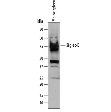
Detection of Mouse Siglec‑E by Western Blot.
Western blot shows lysates of mouse spleen tissue. PVDF membrane was probed with 1 µg/mL of Goat Anti-Mouse Siglec-E Antigen Affinity-purified Polyclonal Antibody (Catalog # AF5806) followed by HRP-conjugated Anti-Goat IgG Secondary Antibody (Catalog # HAF109). A specific band was detected for Siglec-E at approximately 80 kDa (as indicated). This experiment was conducted under reducing conditions and using Immunoblot Buffer Group 1.Siglec‑E in Mouse Spleen.
Siglec-E was detected in perfusion fixed frozen sections of mouse spleen using Goat Anti-Mouse Siglec-E Antigen Affinity-purified Polyclonal Antibody (Catalog # AF5806) at 1.7 µg/mL overnight at 4 °C. Tissue was stained using the NorthernLights™ 557-conjugated Anti-Goat IgG Secondary Antibody (red; Catalog # NL001) and counterstained with DAPI (blue). Specific staining was localized to plasma membrane. View our protocol for Fluorescent IHC Staining of Frozen Tissue Sections.Applications for Mouse Siglec-E Antibody
Application
Recommended Usage
Immunohistochemistry
5-15 µg/mL
Sample: Perfusion fixed frozen sections of mouse spleen
Sample: Perfusion fixed frozen sections of mouse spleen
Western Blot
1 µg/mL
Sample: Mouse spleen tissue
Sample: Mouse spleen tissue
Formulation, Preparation, and Storage
Purification
Antigen Affinity-purified
Reconstitution
Reconstitute at 0.2 mg/mL in sterile PBS. For liquid material, refer to CoA for concentration.
Formulation
Lyophilized from a 0.2 μm filtered solution in PBS with Trehalose. *Small pack size (SP) is supplied either lyophilized or as a 0.2 µm filtered solution in PBS.
Shipping
Lyophilized product is shipped at ambient temperature. Liquid small pack size (-SP) is shipped with polar packs. Upon receipt, store immediately at the temperature recommended below.
Stability & Storage
Use a manual defrost freezer and avoid repeated freeze-thaw cycles.
- 12 months from date of receipt, -20 to -70 °C as supplied.
- 1 month, 2 to 8 °C under sterile conditions after reconstitution.
- 6 months, -20 to -70 °C under sterile conditions after reconstitution.
Background: Siglec-E
References
- Varki, A. and T. Angata (2006) Glycobiology 16:1R.
- Crocker, P.R. et al. (2007) Nat. Rev. Immunol. 7:255.
- Yu, Z. et al. (2001) Biochem. J. 353:483.
- Ulyanova, T. et al. (2001) J. Biol. Chem. 276:14451.
- Zhang, J.Q. et al. (2004) Eur. J. Immunol. 34:1175.
- Boyd, C.R. et al. (2009) J. Immunol. 183:7703.
- Erdmann, H. et al. (2009) Cell. Microbiol. 11:1600.
Long Name
Sialic Acid Binding Ig-like Lectin E
Alternate Names
SiglecE
Entrez Gene IDs
83382 (Mouse)
Gene Symbol
SIGLECE
UniProt
Additional Siglec-E Products
Product Documents for Mouse Siglec-E Antibody
Product Specific Notices for Mouse Siglec-E Antibody
For research use only
Loading...
Loading...
Loading...
Loading...

