Mouse Spi-B Antibody
R&D Systems, part of Bio-Techne | Catalog # AF7204

Key Product Details
Validated by
Species Reactivity
Validated:
Cited:
Applications
Validated:
Cited:
Label
Antibody Source
Product Specifications
Immunogen
Tyr18-Glu167 (Tyr110Phe)
Accession # O35906
Specificity
Clonality
Host
Isotype
Scientific Data Images for Mouse Spi-B Antibody
Spi-B in Mouse Splenocytes.
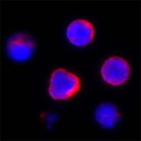
Spi-B was detected in immersion fixed mouse splenocytes using Sheep Anti-Mouse Spi-B Antigen Affinity-purified Polyclonal Antibody (Catalog # AF7204) at 15 µg/mL for 3 hours at room temperature. Cells were stained using the NorthernLights™ 557-conjugated Anti-Sheep IgG Secondary Antibody (red; Catalog # NL010) and counterstained with DAPI (blue). Specific staining was localized to plasma membranes and cytoplasm. View our protocol for Fluorescent ICC Staining of Non-adherent Cells.Detection of SPi-B in Mouse Splenocytes by Flow Cytometry.
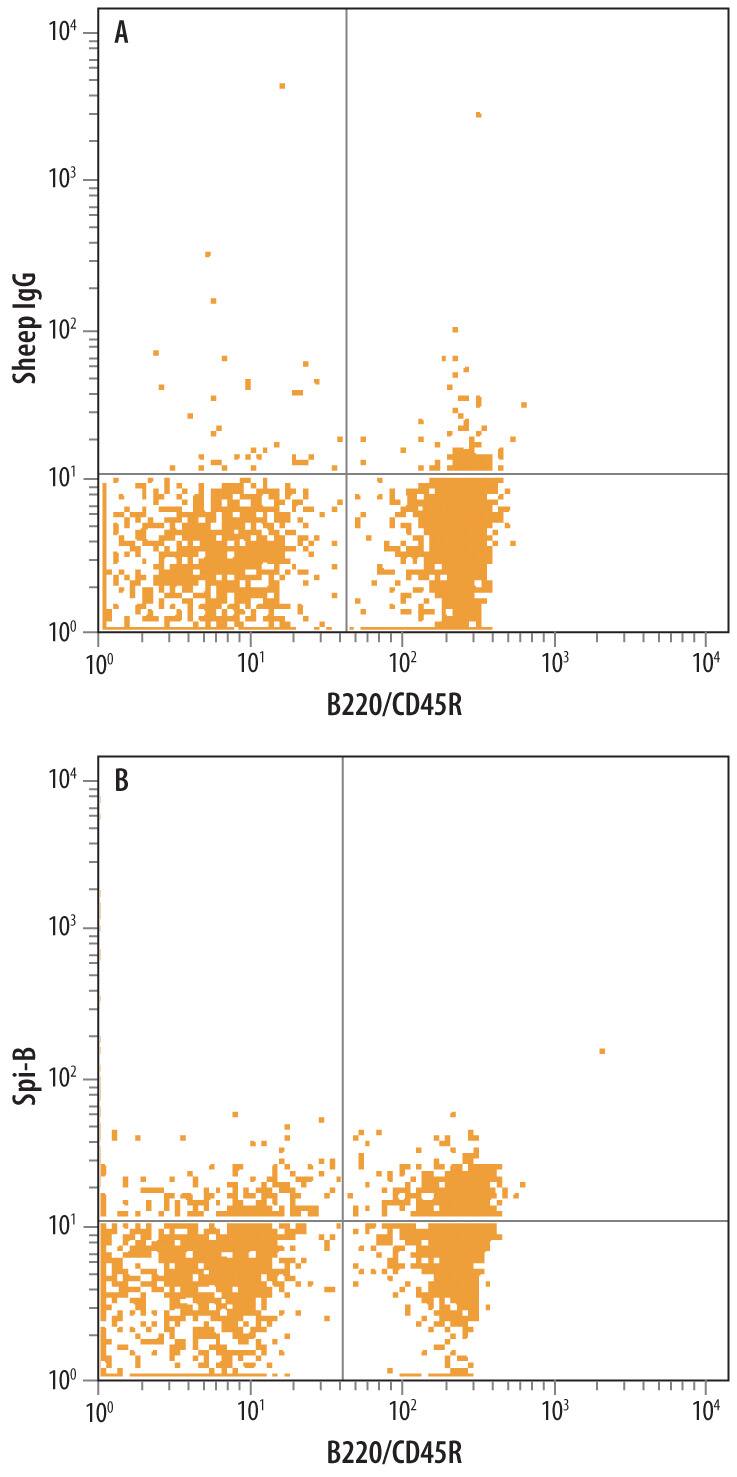
Mouse splenocytes were stained with Sheep Anti-Mouse Spi-B Antigen Affinity-purified Polyclonal Antibody (Catalog # AF7204) followed by Allophycocyanin-conjugated Anti-Sheep IgG Secondary Antibody (Catalog # F0127) and Rat Anti-Mouse B220/CD45R PE-conjugated Monoclonal Antibody (Catalog # FAB1217P). Quadrant markers were set based on control antibody staining (Catalog # 5-001-A). To facilitate intracellular staining, cells were fixed with paraformadehyde and permeabilized with saponin.Detection of Mouse Spi-B by Immunocytochemistry/Immunofluorescence
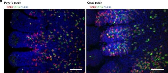
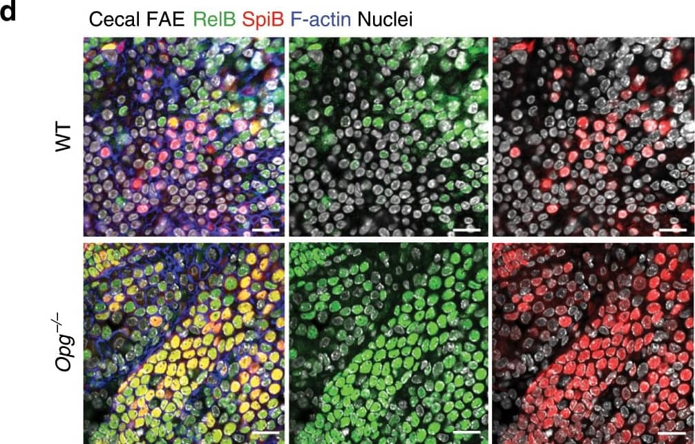
OPGhigh M cells cluster more in cecal patches than in Peyer’s patches.a Whole-mount immunostaining of the FAE of Peyer’s patches (left) and cecal patches (right) for OPG (green) and Spi-B (red). Nuclei were stained with DAPI (blue); scale bars: 50 µm. b Scatter plots of the fluorescence intensities of OPG versus Spi-B. Red dots represent cells stained with anti-Spi-B antibody that was conjugated with HyLyte Fluor 555 and anti-OPG antibody that was conjugated with HyLyte Fluor 647. Blue dots represent background fluorescence intensity of randomly selected non-stained cells. Fluorescence intensities were measured for at least 3000 cells from five FAEs of three mice. c Frequencies of OPGhigh M cells in Peyer’s patches and cecal patches were quantified. *p < 0.05, ***p < 0.005. Student’s t-test, n = 5 FAE from three animals. The source data underlying panels b and c are provided as a Source Data file. Image collected and cropped by CiteAb from the following publication (https://pubmed.ncbi.nlm.nih.gov/31932605), licensed under a CC-BY license. Not internally tested by R&D Systems.Applications for Mouse Spi-B Antibody
CyTOF-ready
Immunocytochemistry
Sample: Immersion fixed mouse splenocytes
Intracellular Staining by Flow Cytometry
Sample: Mouse splenocytes fixed with paraformadehyde and permeabilized with saponin
Formulation, Preparation, and Storage
Purification
Reconstitution
Formulation
Shipping
Stability & Storage
- 12 months from date of receipt, -20 to -70 °C as supplied.
- 1 month, 2 to 8 °C under sterile conditions after reconstitution.
- 6 months, -20 to -70 °C under sterile conditions after reconstitution.
Background: Spi-B
Spi-B (Transcription factor Spi-B) is a 33-45 kDa member of the ets family of transcription factors. It is found in hematopoietic cells such as B cells and plasmacytoid dendritic cells (DC). In transitional B cells, Spi-B promotes their differentiation into follicular (naïve) B cells. In hematopoietic stem cells, Spi-B stimulates the generation of IFN-producing plasmacytoid DC at the expense of T, B and NK cell development. Mouse Spi-B is 267 amino acids (aa) in length. It contains a dual transactivation region (aa 1-62), plus a PEST domain (aa 110-170) and an Ets DNA-binding domain (aa 174-257). There are two isoform variants. One shows a nine aa substitution for aa 1-8, while a second possesses an 18 aa insertion after Leu17. Over aa 18-167, mouse Spi-B shares 91% and 74% aa identity with rat and human Spi-B, respectively.
Long Name
Alternate Names
Gene Symbol
UniProt
Additional Spi-B Products
Product Documents for Mouse Spi-B Antibody
Product Specific Notices for Mouse Spi-B Antibody
For research use only


