Mouse TREM2 Biotinylated Antibody
R&D Systems, part of Bio-Techne | Catalog # BAF1729


Key Product Details
Species Reactivity
Validated:
Cited:
Applications
Validated:
Cited:
Label
Antibody Source
Product Specifications
Immunogen
Leu19-Pro168
Accession # Q99NH8
Specificity
Clonality
Host
Isotype
Scientific Data Images for Mouse TREM2 Biotinylated Antibody
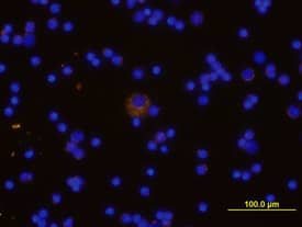
TREM‑2 in Mouse Splenocytes.
TREM-2 was detected in immersion fixed mouse spleno-cytes using Mouse Anti-TREM-2 Biotinylated Antigen Affinity-purified Polyclonal Antibody (Catalog # BAF1729) at 10 µg/mL for 3 hours at room temperature. Cells were stained using the NorthernLights™ 557-conjugated Streptavidin (yellow; Catalog # NL999) and counterstained with DAPI (blue). View our protocol for Fluorescent ICC Staining of Non-adherent Cells.Mouse TREM2 ELISA Standard Curve.
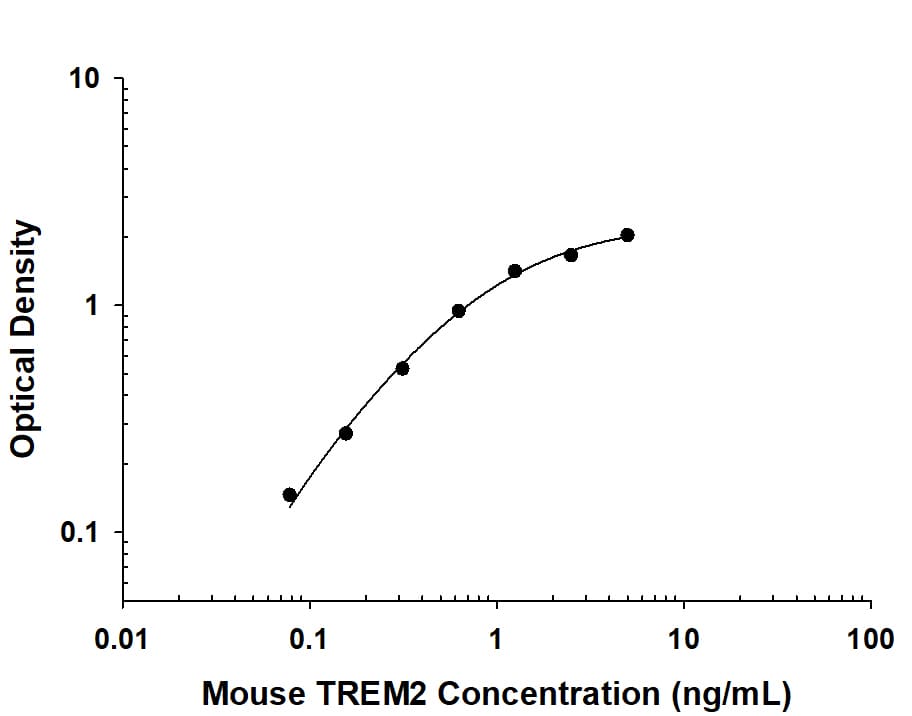
Recombinant Human/Mouse TREM2 protein was serially diluted 2-fold and captured by Rat Anti-Human/Mouse TREM2 Monoclonal Antibody (Catalog # MAB17291) coated on a Clear Polystyrene Microplate (Catalog # DY990). Sheep Anti-Mouse TREM2 Biotinylated Antigen Affinity-purified Polyclonal Antibody (Catalog # BAF1729) was incubated with the protein captured on the plate. Detection of the standard curve was achieved by incubating Streptavidin-HRP (Catalog # DY998) followed by Substrate Solution (Catalog # DY999) and stopping the enzymatic reaction with Stop Solution (Catalog # DY994).Mouse TREM2 ELISA Standard Curve.
Recombinant Mouse TREM2 protein was serially diluted 2-fold and captured by Sheep Anti-Mouse TREM2 Antigen Affinity-purified Polyclonal Antibody (Catalog # AF1729) coated on a Clear Polystyrene Microplate (Catalog # DY990). Sheep Anti-Mouse TREM2 Biotinylated Antigen Affinity-purified Polyclonal Antibody (Catalog # BAF1729) was incubated with the protein captured on the plate. Detection of the standard curve was achieved by incubating Streptavidin-HRP (Catalog # DY998) followed by Substrate Solution (Catalog # DY999) and stopping the enzymatic reaction with Stop Solution (Catalog # DY994).Applications for Mouse TREM2 Biotinylated Antibody
ELISA
This antibody functions as an ELISA detection antibody when paired with Rat Anti-Human/Mouse TREM2 Monoclonal Antibody (Catalog # MAB17291) or Sheep Anti-Mouse TREM2 Antigen Affinity-purified Polyclonal Antibody (Catalog # AF1729)
This product is intended for assay development on various assay platforms requiring antibody pairs.
Immunocytochemistry
Sample: Immersion fixed mouse splenocytes
Western Blot
Sample: Recombinant Mouse TREM2b Fc Chimera (Catalog # 1729-T2)
Formulation, Preparation, and Storage
Purification
Reconstitution
Formulation
Shipping
Stability & Storage
- 12 months from date of receipt, -20 to -70 °C as supplied.
- 1 month, 2 to 8 °C under sterile conditions after reconstitution.
- 6 months, -20 to -70 °C under sterile conditions after reconstitution.
Background: TREM2
TREM2 (Triggering Receptor Expressed by Myeloid cells) is an Ig superfamily cell surface receptor that activates a number of myeloid cell types (1). It is a member of a small gene family located on human chromosome 6p21 and mouse chromosome 17 in a region linked to the MHC (2). A single human TREM2 gene has been described, however, two closely related orthologs were reported in mouse (3). The proteins differ by only three amino acids and were designated TREM2a and TREM2b. TREM2 is type I transmembrane protein consisting of a single extracellular immunoglobulin (V-like) domain, a transmembrane domain with a positively charged lysine residue, and a short cytoplasmic tail (1). It associates with the signal adapter protein, DAP12, for signaling and function. DAP12 has a cytoplasmic ITAM that is phosphorylated upon ligand binding leading to the subsequent activation of cytoplasmic tyrosine kinases. TREM2 is expressed by immature monocyte-derived dendritic cells (DC), and expression is down-regulated upon activation of DC by microbial products and costimulatory signals (4). Ligation of TREM2 on immature DC with anti-TREM2 antibodies results in partial DC activation and the up-regulation of CCR7 and some co-stimulatory molecules. A role for TREM2 in the functioning of osteoclasts and microglia is suggested by the discovery that homozygous loss-of-function mutations in either TREM2 or DAP12 result in Nasu-Hakola disease characterized by a combination of presenile demetia and bone cysts (5). In vitro studies indicate that the differentiation of myeloid precursors into osteoclasts is dramatically impaired in TREM2 deficient individuals (6).
References
- Colonna, M. (2003) Nature Rev. Immunol. 3:445.
- Allcock, R. et al. (2003) Eur. J. Immunol. 33:567.
- Daws, M. et al. (2001) Eur. J. Immunol. 31:783.
- Bouchon, A. et al. (2001) J. Exp. Med. 194:1111.
- Paloneva, J. et al. (2002) Am. J. Hum. Genet. 71:656.
- Cella, M. et al. (2003) J. Exp. Med. 198:645.
Long Name
Alternate Names
Gene Symbol
UniProt
Additional TREM2 Products
Product Documents for Mouse TREM2 Biotinylated Antibody
Product Specific Notices for Mouse TREM2 Biotinylated Antibody
For research use only

