Mouse VISTA/B7-H5/PD-1H Antibody
R&D Systems, part of Bio-Techne | Catalog # AF7005


Key Product Details
Species Reactivity
Applications
Label
Antibody Source
Product Specifications
Immunogen
Phe33-Ala191
Accession # Q9D659
Specificity
Clonality
Host
Isotype
Scientific Data Images for Mouse VISTA/B7-H5/PD-1H Antibody
Detection of VISTA/B7-H5/PD-1H by Western Blot.
Western blot shows lysates of mouse platelets. PVDF membrane was probed with 1 µg/mL of Sheep Anti-Mouse VISTA/B7-H5/PD-1H Antigen Affinity-purified Polyclonal Antibody (Catalog # AF7005) followed by HRP-conjugated Anti-Sheep IgG Secondary Antibody (Catalog # HAF016). A specific band was detected for VISTA/B7-H5/PD-1H at approximately 30 kDa (as indicated). This experiment was conducted under reducing conditions and using Immunoblot Buffer Group 8.Detection of VISTA/B7-H5/PD-1H in HEK293 Human Cell Line Transfected with Mouse VISTA and eGFP by Flow Cytometry.
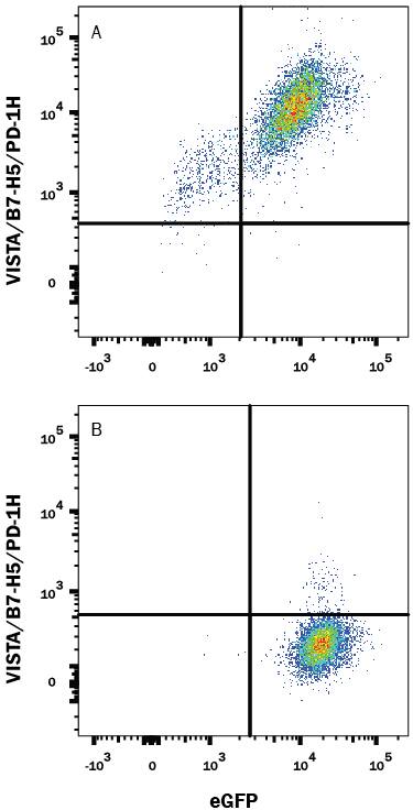
HEK293 human embryonic kidney cell line transfected with either (A) mouse VISTA or (B) irrelevant transfectants and eGFP was stained with Sheep Anti-Mouse VISTA/B7-H5/PD-1H Antigen Affinity-purified Polyclonal Antibody (Catalog # AF7005) followed by Allophycocyanin-conjugated Anti-Sheep IgG Secondary Antibody (Catalog # F0127). Quadrant markers were set based on control antibody staining (Catalog # 5-001-A). View our protocol for Staining Membrane-associated Proteins.VISTA/B7-H5/PD-1H in D3 Mouse Cell Line.
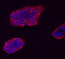
VISTA/B7-H5/PD-1H was detected in immersion fixed D3 mouse embryonic stem cell line using Sheep Anti-Mouse VISTA/B7-H5/PD-1H Antigen Affinity-purified Polyclonal Antibody (Catalog # AF7005) at 10 µg/mL for 3 hours at room temperature. Cells were stained using the NorthernLights™ 557-conjugated Anti-Sheep IgG Secondary Antibody (red; Catalog # NL010) and counterstained with DAPI (blue). Specific staining was localized to cell surfaces and cytoplasm. View our protocol for Fluorescent ICC Staining of Cells on Coverslips.Applications for Mouse VISTA/B7-H5/PD-1H Antibody
CyTOF-ready
Flow Cytometry
Sample: HEK293 human embryonic kidney cell line transfected with mouse VISTA and eGFP
Immunocytochemistry
Sample: Immersion fixed D3 mouse embryonic stem cell line
Western Blot
Sample: Mouse platelets
Formulation, Preparation, and Storage
Purification
Reconstitution
Formulation
Shipping
Stability & Storage
- 12 months from date of receipt, -20 to -70 °C as supplied.
- 1 month, 2 to 8 °C under sterile conditions after reconstitution.
- 6 months, -20 to -70 °C under sterile conditions after reconstitution.
Background: VISTA/B7-H5/PD-1H
Platelet Receptor Gi24 (also known as VISTA, B7-H5, SISP1, C10orf54 and , Dies1 [Differentiation of ESC-1]) is a 55-65 kDa member of the Ig superfamily. It is a transmembrane molecule expressed in bone, on embryonic stem cells (ESCs), and on tumor cell surfaces. On ESCs, Gi24 appears to positively interact with BMP4, potentiating BMP signaling and the transition from an undifferentiated to a differentiated state. On tumor cells, Gi24 both promotes MT1-MMP expression and activity, and serves as a substrate for MT1-MMP. This increases the potential for cell motility. Mature mouse Gi24 is a 277 amino acid (aa) type I transmembrane glycoprotein (aa 33-309). It contains a 149 aa extracellular region (aa 33-191) with one V-type Ig-like domain (aa 33-161) and a 97 aa cytoplasmic domain. Based on human Gi24, mouse Gi24 will likely undergo proteolytic cleavage by MT1-MMP, generating a soluble 30 kDa extracellular fragment, plus a 25-30 kDa membrane-bound fragment. There are two potential isoform variants. One contains a deletion of aa 127-187, while another shows an alternative start site at Met82. Over aa 33-191, mouse Gi24/Dies1 shares 78% and 70% aa identity with rat and human Gi24, respectively.
Alternate Names
Gene Symbol
UniProt
Additional VISTA/B7-H5/PD-1H Products
Product Documents for Mouse VISTA/B7-H5/PD-1H Antibody
Product Specific Notices for Mouse VISTA/B7-H5/PD-1H Antibody
For research use only

