Mouse VSIG4 Antibody
R&D Systems, part of Bio-Techne | Catalog # AF4674

Key Product Details
Species Reactivity
Validated:
Cited:
Applications
Validated:
Cited:
Label
Antibody Source
Product Specifications
Immunogen
His20-Pro187
Accession # NP_808457
Specificity
Clonality
Host
Isotype
Scientific Data Images for Mouse VSIG4 Antibody
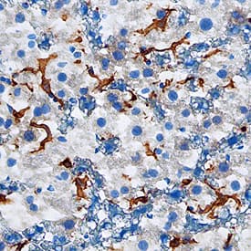
VSIG4 in Mouse Liver.
VSIG4 was detected in perfusion fixed frozen sections of mouse liver using Goat Anti-Mouse VSIG4 Antigen Affinity-purified Polyclonal Antibody (Catalog # AF4674) at 1.7 µg/mL overnight at 4 °C. Tissue was stained using the Anti-Goat HRP-DAB Cell & Tissue Staining Kit (brown; Catalog # CTS008) and counterstained with hematoxylin (blue). Specific staining was localized to Kuppfer cells. View our protocol for Chromogenic IHC Staining of Frozen Tissue Sections.Applications for Mouse VSIG4 Antibody
Immunohistochemistry
Sample: Perfusion fixed frozen sections of mouse liver
Formulation, Preparation, and Storage
Purification
Reconstitution
Formulation
Shipping
Stability & Storage
- 12 months from date of receipt, -20 to -70 °C as supplied.
- 1 month, 2 to 8 °C under sterile conditions after reconstitution.
- 6 months, -20 to -70 °C under sterile conditions after reconstitution.
Background: VSIG4
Mouse VSIG4 (V-set and immunoglobulin domain containing 4), also known as CRIg and Z39IG, is a type I transmembrane glycoprotein that is a B7 family-related protein and an Ig superfamily member (1‑2). Mouse VSIG4 is synthesized as a 280 amino acid (aa) precursor that contains a signal sequence, an IgV-type immunological domain (aa 36‑115), one potential N-linked glycosylation site, and a single transmembrane domain (2). The IgV domain of mouse VSIG4 shares 86% and 80% aa sequence identity with the IgV domains of rat and human VSIG4, respectively. Quantitative PCR reveals that VSIG4 mRNA is expressed at high levels in the liver, dendritic cells, neutrophils, and macrophages, and at lower levels in the lung, heart, spleen, and lymph nodes (1). No VSIG4 expression appears to be present in T and B cells (1). The use of polyclonal rabbit serum against the extracellular domain of VSIG4 demonstrates that VSIG4 is specifically expressed on naïve resting tissue macrophages, and that the expression is downregulated or lost upon activation (1). Furthermore, histological analysis shows expression of VSIG4+ macrophages only in the liver, thymic medulla, and heart (1). VSIG4 macrophages are not detected in the intestines, kidney, skeletal muscle, lymph node, splenic white pulp, lung, or brain (1). Studies show that VSIG4/Fc Chimera strongly inhibits proliferation of anti-CD3 as well as anti-CD3/anti-CD28-stimulated T cells (1). Indeed, VSIG4 functions as a negative regulator of mouse as well as human T cell activation, and may be involved in the maintenance of peripheral T cell tolerance and/or unresponsiveness (1). In addition, VSIG4’s expression on Kupffer cells is required for efficient binding and phagocytosis of complement C3 opsonized particles (2). VSIG4 acts as a macrophage complement receptor by binding complement fragments C3b and iC3b (2). VSIG4 binding to C3b inhibits complement activation through the alternative pathway, making it a potent suppressor of established inflammation (3‑4).
References
- Vogt, L. et al. (2006) J. Clin. Invest. 116:2817.
- Helmy, K. et al. (2006) Cell 124:915.
- Katschke, K.J. et al. (2007) J. Exp. Med. 204:1319.
- Wiesmann, C. et al. (2006) Nature 444:217.
Long Name
Alternate Names
Gene Symbol
UniProt
Additional VSIG4 Products
Product Documents for Mouse VSIG4 Antibody
Product Specific Notices for Mouse VSIG4 Antibody
For research use only
