Western Blotting of NLRP3/NALP3 in Multiple Species
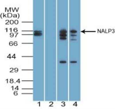
Analysis of NALP3 using NALP3 antibody. Human testis lysate in the 1) absence, 2) presence of immunizing peptide, 3) mouse and 4) rat testis probed with NALP3 antibody at 5, 2 and 2 ug/mL respectively. Goat anti-rabbit IgG HRP secondary antibody and PicoTect ECL substrate solution were used for this test.
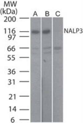
Western Blot Detection of NLRP3/NALP3 in Multiple Cell Lysates
Analysis of NALP3 using NALP3 antibody. Lane A) Human NALP3 transfected cell lysate, B) Mouse NALP3 transfected cell lysate, and C) HEK293 control lysate probed with NALP3 antibody at 3 ug/mL.
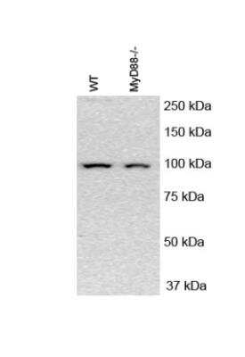
Western Blot Analysis of NLRP3/NALP3 in Mouse Cell Lysate
Analysis in mouse cell lysate.
Western Blot Detection of NLRP3/NALP3 in HFD Rats
Regular aerobic exercise decreased myocardial inflammation in HFD rats. Representative Western blot analysis of NLRP3 and caspase-1. Image collected and cropped by CiteAb from the following publication (https://www.frontiersin.org/article/10.3389/fphys.2019.01286/full), licensed under a CC-BY license.
Western Blot: NLRP3/NALP3 Antibody - BSA Free [NBP2-12446] -
Effect of Taohong Siwu decoction (THSWD) on the characteristic protein of pyroptosis in middle cerebral artery occlusion-reperfusion (MCAO/R) rats. (A) Photographs of western blots, (B) NLRP3, (C) Caspase-1, (D) Caspase-1 p10, (E) ASC, (F) GSDMD. a: Sham, b: Model, c: THSWD (18 g/kg), d: THSWD (9 g/kg), e: THSWD (4.5 g/kg), f: nimodipine. The results were presented as the mean ± SD (n = 3). Compared with sham group, #p < 0.05, ##p < 0.01. Compared with model group, *p < 0.05, **p < 0.01.
Western Blot: NLRP3/NALP3 Antibody - BSA Free [NBP2-12446] -
Western Blot: NLRP3/NALP3 Antibody - BSA Free [NBP2-12446] - Downstream receptor for advanced glycation end-products (RAGE) & nuclear factor (NF)-kappa B pathway were inhibited after treatment of Nec-1 & melatonin. NF-kappa B pathway was detected by immunoblotting in the cortex (A–E) & hippocampus CA1 (F–J). Relative downstream inflammatory factors were detected by immunoblotting in the cortex (K–N) & hippocampus CA1 (O–R). All experiments were performed in triplicate by one way ANOVA plus Tukey’s test. **P < 0.01 & ***P < 0.001 vs. sham group. #P < 0.05 & ##P < 0.01 vs. CCI group. Image collected & cropped by CiteAb from the following publication (https://pubmed.ncbi.nlm.nih.gov/31607859), licensed under a CC-BY license. Not internally tested by Novus Biologicals.
Immunocytochemistry/ Immunofluorescence: NLRP3/NALP3 Antibody - BSA Free [NBP2-12446] -
Immunocytochemistry/ Immunofluorescence: NLRP3/NALP3 Antibody - BSA Free [NBP2-12446] - Immunofluorescence staining of TXNIP (a) & NLRP3 (b). NG: normal glucose (5.6 mM); HG: high glucose (30 mM). NH/R: hypoxia (4 h)/reoxygenation (2 h) under NG conditions; HH/R: hypoxia (4 h)/reoxygenation (2 h) under HG conditions. HH/R-RES: HH/R pretreated by RES (50 μM) for 72 h with the high glucose incubation. HH/R-siRNA: TXNIP protein was inhibited by transfection with TXNIP siRNA before HH/R; HH/R-scrambled siRNA: scrambled siRNA used as control before HH/R. Image collected & cropped by CiteAb from the following publication (https://pubmed.ncbi.nlm.nih.gov/27867451), licensed under a CC-BY license. Not internally tested by Novus Biologicals.
Western Blot: NLRP3/NALP3 Antibody - BSA Free [NBP2-12446] -
Western Blot: NLRP3/NALP3 Antibody - BSA Free [NBP2-12446] - MSC-EVs inhibit activity of NLRP3 inflammasome in SDH of IC rats. A–E Western blot analysis showing that intrathecal injection of MSC-EVs significantly decreased expression levels of NLRP3, Caspase-1, IL-1 beta & IL-18 in SDH of IC rats. n = 8 per group. *P < 0.05 Image collected & cropped by CiteAb from the following publication (https://pubmed.ncbi.nlm.nih.gov/35387668), licensed under a CC-BY license. Not internally tested by Novus Biologicals.
Western Blot: NLRP3/NALP3 Antibody - BSA Free [NBP2-12446] -
Western Blot: NLRP3/NALP3 Antibody - BSA Free [NBP2-12446] - Effect of Taohong Siwu decoction (THSWD) on the characteristic protein of pyroptosis in middle cerebral artery occlusion-reperfusion (MCAO/R) rats. (A) Photographs of western blots, (B) NLRP3, (C) Caspase-1, (D) Caspase-1 p10, (E) ASC, (F) GSDMD. a: Sham, b: Model, c: THSWD (18 g/kg), d: THSWD (9 g/kg), e: THSWD (4.5 g/kg), f: nimodipine. The results were presented as the mean ± SD (n = 3). Compared with sham group, #p < 0.05, ##p < 0.01. Compared with model group, *p < 0.05, **p < 0.01. Image collected & cropped by CiteAb from the following publication (https://pubmed.ncbi.nlm.nih.gov/33424599), licensed under a CC-BY license. Not internally tested by Novus Biologicals.
Western Blot: NLRP3/NALP3 Antibody - BSA Free [NBP2-12446] -
Western Blot: NLRP3/NALP3 Antibody - BSA Free [NBP2-12446] - Hippocampal AMPK/Sirt1 & NF kappaB/NLRP3/IL-1 beta signaling pathways in diabetic rats & the effects of aerobic exercise. Type 2 diabetes significantly decreases the activation of hippocampal AMPK (a) & the level of Sirt1 (c), leading to increased Ac-NF kappaB (d) & NF kappaB (e); diabetic rats contain more NLRP3 inflammasomes (f) & IL-1 beta (g). Aerobic exercise intervention significantly increases AMPK activity & Sirt1 concentration; aerobic exercise downregulates the Ac-NF kappaB & NF kappaB, leading to decreased levels of the NLRP3 inflammasome & IL-1 beta. ∗∗p < 0.01, DM group vs. C group; #p < 0.05, TDM group vs. DM group. Image collected & cropped by CiteAb from the following publication (https://pubmed.ncbi.nlm.nih.gov/30911292), licensed under a CC-BY license. Not internally tested by Novus Biologicals.
Western Blot: NLRP3/NALP3 Antibody - BSA Free [NBP2-12446] -
Western Blot: NLRP3/NALP3 Antibody - BSA Free [NBP2-12446] - The inflammasome inhibitor reduced the myocardial infarct size & decreased activation of the NLRP3 inflammasome, IL-1 beta, & caspase-1 (p10) in nondiabetic & diabetic rats after MI/R. The infarct size was detected by TTC staining (a). The CK-MB activities were determined by enzyme activity assay kits (b). The expression of NLRP3 (c), ASC (d), procaspase-1 & caspase-1 (e), & IL-1 beta (f) was analyzed by Western blot as shown. Data are expressed as the mean ± SD. n = 6 to 8. ∗∗P < 0.01 versus IR. Image collected & cropped by CiteAb from the following publication (https://pubmed.ncbi.nlm.nih.gov/29062465), licensed under a CC-BY license. Not internally tested by Novus Biologicals.
Western Blot: NLRP3/NALP3 Antibody - BSA Free [NBP2-12446] -
Western Blot: NLRP3/NALP3 Antibody - BSA Free [NBP2-12446] - Protein expression in HG & H/R conditions. Western blotting for NLRP3 (a), ASC (b), procaspase-1 & caspase-1 (p10) (c), & IL-1 beta (d) in H9C2 cells. Data are expressed as the mean ± SD. n = 5 per group. ∗∗P < 0.01 versus N; #P < 0.01 versus LG; & ##P < 0.01 versus LG + H/R. Image collected & cropped by CiteAb from the following publication (https://pubmed.ncbi.nlm.nih.gov/29062465), licensed under a CC-BY license. Not internally tested by Novus Biologicals.
Western Blot: NLRP3/NALP3 Antibody - BSA Free [NBP2-12446] -
Western Blot: NLRP3/NALP3 Antibody - BSA Free [NBP2-12446] - SZF inhibited the activation of the NLRP3-ASC-caspase-1 axis by suppressing TXNIP. Gene & protein expression levels in the renal tissue: (a) TXNIP; (b) NLRP3; (c) ASC; (d) caspase-1 & Procaspase-1. Protein levels were determined by Western blotting, were quantified through densitometry, & are expressed as the optical density ratio to GAPDH. mRNA levels were determined through real-time PCR. The numbers in the Western blot figures represent groups: 1 means Control, 2 means OA, 3 means OA + Allopurinol, & 4 means OA + SZF. Data are expressed as the mean ± SD (n = 3). ∗P < 0.05 versus the OA group. #P > 0.05 versus the OA + Allopurinol group. Image collected & cropped by CiteAb from the following publication (https://pubmed.ncbi.nlm.nih.gov/29358971), licensed under a CC-BY license. Not internally tested by Novus Biologicals.
Western Blot: NLRP3/NALP3 Antibody - BSA Free [NBP2-12446] -
Western Blot: NLRP3/NALP3 Antibody - BSA Free [NBP2-12446] - Western blot analysis of TXNIP & NLRP3 protein expression in cultured HK-2 cells treated by normal glucose (5.5 mM), high glucose (30 mM), & NG + mannitol, respectively, for 72 hours, then following 4 hours of hypoxia & 2 hours of reoxygenation in HK-2 cells under high glucose stimulation with or without TXNIP siRNA & RES treatment, respectively. Representative blots (a) & quantitative analysis of Western blots for TXNIP (c) & NLRP3 (d), activity of caspase-1 (e), level of IL-1 beta (f), & Western blot of TXNIP gene knockdown in HK-2 cells (b). The data in (c–f) are means ± SE (n = 5). #P < 0.05 versus NG group; ☆P < 0.05 versus NH/R group; &P < 0.05 versus HH/R-scrambled siRNA group. Image collected & cropped by CiteAb from the following publication (https://pubmed.ncbi.nlm.nih.gov/27867451), licensed under a CC-BY license. Not internally tested by Novus Biologicals.
Immunohistochemistry: NLRP3/NALP3 Antibody - BSA Free [NBP2-12446] -
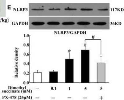
Immunohistochemistry: NLRP3/NALP3 Antibody - BSA Free [NBP2-12446] - C-AR inhibited NLRP3 inflammasome activation in synovial tissue. (A,B) Immunohistochemistry staining of NLRP3 expression (A) & cleaved caspase-1 activation (B) in the synovial tissue of collagen-induced arthritis (CIA) rats (original magnification, 100 × ; scale bar, 50 μm). (C,D) IL-1 beta in the synovial tissue of CIA rats (C) & TGF-beta 1-stimulated synovial fibroblasts (D) were measured by ELISA Kits (n = 6); (E) NLRP3 protein expression in succinate-stimulated synovial fibroblasts. The results were derived from four independent experiments for immunohistochemistry staining & Western blot & expressed as the mean ± SD. *p < 0.05 vs. the model; #p < 0.05 vs. the indicated treatment. Image collected & cropped by CiteAb from the following publication (http://journal.frontiersin.org/article/10.3389/fimmu.2016.00532/full), licensed under a CC-BY license. Not internally tested by Novus Biologicals.
Western Blot: NLRP3/NALP3 Antibody - BSA Free [NBP2-12446] -
Western Blot: NLRP3/NALP3 Antibody - BSA Free [NBP2-12446] - The ROS scavenger inhibited the activation of NLRP3 inflammasomes in H9C2 cells exposed to H/R injury. The expression levels of NLRP3 (a), ASC (b), procaspase-1 & caspase-1 (p10) (c), & IL-1 beta (d) were detected by Western blot. Data are expressed as the mean ± SD. n = 5 per group. ∗P < 0.05 & ∗∗P < 0.01 versus LG + H/R; #P < 0.05 & ##P < 0.01 versus HG + H/R. Image collected & cropped by CiteAb from the following publication (https://pubmed.ncbi.nlm.nih.gov/29062465), licensed under a CC-BY license. Not internally tested by Novus Biologicals.
Western Blot: NLRP3/NALP3 Antibody - BSA Free [NBP2-12446] -
Western Blot: NLRP3/NALP3 Antibody - BSA Free [NBP2-12446] - The activation of the NLRP3 inflammasome & expression of caspase-1 & IL-1 beta were increased in diabetic rats after MI/R insult. NLRP3 & caspase-1 expression in heart tissues were examined by immunohistochemistry (a). The expression of NLRP3 (b), ASC (c), procaspase-1 & caspase-1 (d), & IL-1 beta (e) were analyzed by Western blot. Data are expressed as the mean ± SD. n = 8. ∗∗P < 0.01 versus sham; #P < 0.05 versus Ctrl + sham; & ##P < 0.01 versus Ctrl + I/R. Image collected & cropped by CiteAb from the following publication (https://pubmed.ncbi.nlm.nih.gov/29062465), licensed under a CC-BY license. Not internally tested by Novus Biologicals.
Western Blot: NLRP3/NALP3 Antibody - BSA Free [NBP2-12446] -
Western Blot: NLRP3/NALP3 Antibody - BSA Free [NBP2-12446] - NLRP3 inflammasome is identified to be significantly upregulated in ipsilateral SCDH of CPIP rats. a Heat map showing the expression of NLR family genes identified in ipsilateral SCDH of CPIP rats vs. sham rats. n = 3 rats/group. b–d qPCR validation of the upregulation of Nlrp3 (b), Caspase-1 (c), & Il-1 beta (d) genes in ipsilateral SCDH of CPIP rats vs. sham rats. n = 8 rats/group. e–g Western blot analysis of NLRP3 (e), Caspase-1 (f), & IL-1 beta (g) protein expressions in ipsilateral SCDH of CPIP rats vs. sham rats. n = 8 rats/group. *p < 0.05, **p < 0.01 vs. sham group. Student’s t test was used for comparisons Image collected & cropped by CiteAb from the following publication (https://pubmed.ncbi.nlm.nih.gov/32446302), licensed under a CC-BY license. Not internally tested by Novus Biologicals.
Immunohistochemistry: NLRP3/NALP3 Antibody - BSA Free [NBP2-12446] -
Immunohistochemistry: NLRP3/NALP3 Antibody - BSA Free [NBP2-12446] - Exendin-4 inhibits diabetic cardiomyocyte pyroptosis: (a) immunohistochemistry of NLRP3, cleaved caspase-1, IL-1 beta, & IL-18 in the myocardium; (b, c) transcriptional activity of caspase-1 & NLRP3 in the heart (n = 3); (d, e) IL-1 beta & IL-18 ELISA with supernatant of cardiomyocyte culture medium; (f) Western blot of pyroptotic proteins; (g) caspase-1 activity assay (n = 3); (h) FLICA staining in cardiomyocytes. Upper panels showed the fluorescent images of CON, HG, & HG+EXE cardiomyocytes. The lower panel is a statistic of the enrichment of FLICA emitting fluorescence in single cells (n = 10). Image collected & cropped by CiteAb from the following publication (https://pubmed.ncbi.nlm.nih.gov/31886288), licensed under a CC-BY license. Not internally tested by Novus Biologicals.
Western Blot: NLRP3/NALP3 Antibody - BSA Free [NBP2-12446] -
Western Blot: NLRP3/NALP3 Antibody - BSA Free [NBP2-12446] - NLRP3 inflammasome is activated in neurons in SDH of IC rats, & MCC950 inhibits the NLRP3 inflammasome activation. A–E Western blot analysis showing that expression levels of NLRP3, Caspase-1, IL-1 beta & IL-18 were significantly increased in SDH of IC rats compared with normal rats, & MCC950 treatment significantly decreased expression levels of NLRP3, Caspase-1, IL-1 beta & IL-18 in SDH of IC rats. n = 8 per group. *P < 0.05. F Immunofluorescence co-staining showing that NLRP3 was colocalized predominantly with NeuN (neuron marker), but scarcely with GFAP (astrocyte marker) or OX-42 (microglia marker) in the SDH. Scale bars = 200 μm Image collected & cropped by CiteAb from the following publication (https://pubmed.ncbi.nlm.nih.gov/35387668), licensed under a CC-BY license. Not internally tested by Novus Biologicals.
Western Blot: NLRP3/NALP3 Antibody - BSA Free [NBP2-12446] -
Western Blot: NLRP3/NALP3 Antibody - BSA Free [NBP2-12446] - The inflammasome inhibitor reduced the myocardial infarct size & decreased activation of the NLRP3 inflammasome, IL-1 beta, & caspase-1 (p10) in nondiabetic & diabetic rats after MI/R. The infarct size was detected by TTC staining (a). The CK-MB activities were determined by enzyme activity assay kits (b). The expression of NLRP3 (c), ASC (d), procaspase-1 & caspase-1 (e), & IL-1 beta (f) was analyzed by Western blot as shown. Data are expressed as the mean ± SD. n = 6 to 8. ∗∗P < 0.01 versus IR. Image collected & cropped by CiteAb from the following publication (https://pubmed.ncbi.nlm.nih.gov/29062465), licensed under a CC-BY license. Not internally tested by Novus Biologicals.
Immunohistochemistry-Paraffin: NLRP3/NALP3 Antibody - BSA Free [NBP2-12446] -
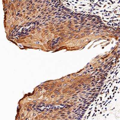
Immunohistochemistry-Paraffin: NLRP3/NALP3 Antibody - BSA Free [NBP2-12446] - The activation of the NLRP3 inflammasome & expression of caspase-1 & IL-1 beta were increased in diabetic rats after MI/R insult. NLRP3 & caspase-1 expression in heart tissues were examined by immunohistochemistry (a). The expression of NLRP3 (b), ASC (c), procaspase-1 & caspase-1 (d), & IL-1 beta (e) were analyzed by Western blot. Data are expressed as the mean ± SD. n = 8. ∗∗P < 0.01 versus sham; #P < 0.05 versus Ctrl + sham; & ##P < 0.01 versus Ctrl + I/R. Image collected & cropped by CiteAb from the following publication (https://pubmed.ncbi.nlm.nih.gov/29062465), licensed under a CC-BY license. Not internally tested by Novus Biologicals.
Western Blot: NLRP3/NALP3 Antibody - BSA Free [NBP2-12446] -
Western Blot: NLRP3/NALP3 Antibody - BSA Free [NBP2-12446] - Exendin-4 inhibits diabetic cardiomyocyte pyroptosis: (a) immunohistochemistry of NLRP3, cleaved caspase-1, IL-1 beta, & IL-18 in the myocardium; (b, c) transcriptional activity of caspase-1 & NLRP3 in the heart (n = 3); (d, e) IL-1 beta & IL-18 ELISA with supernatant of cardiomyocyte culture medium; (f) Western blot of pyroptotic proteins; (g) caspase-1 activity assay (n = 3); (h) FLICA staining in cardiomyocytes. Upper panels showed the fluorescent images of CON, HG, & HG+EXE cardiomyocytes. The lower panel is a statistic of the enrichment of FLICA emitting fluorescence in single cells (n = 10). Image collected & cropped by CiteAb from the following publication (https://pubmed.ncbi.nlm.nih.gov/31886288), licensed under a CC-BY license. Not internally tested by Novus Biologicals.
Western Blot: NLRP3/NALP3 Antibody - BSA Free [NBP2-12446] -
Western Blot: NLRP3/NALP3 Antibody - BSA Free [NBP2-12446] - Glyburide attenuates NLRP3 inflammasome-mediated pyroptosis in HCECs infected with HKCA. HCECs were pretreated with potassium (K+) channel inhibitor (glyburide) for 2 h, & then were incubated with HKCA (MOI = 20) for 24 h. (A,B) Western blot showing the protein levels of NLRP3 in HCECs treated with various concentrations of glyburide (50, 100 & 200 μM) (n = 3). (C,D) Glyburide treatment (200 μM) suppressed the levels of pyroptosis-related proteins (ASC, cleaved CASP1, N-GSDMD, cleaved IL-1 beta & cleaved IL-18) in HCECs challenged with HKCA at 20:1 for 24 h (n = 3). (E) Immunofluorescence analysis of NLRP3, CASP1 & ASC in HCECs pretreated with or without glyburide (200 μM) for 24 h (n = 3). Scale bar = 20 μm; magnification 400×. (F) LDH release of HCECs treated with glyburide (200 μM) (n = 6). CASP1: caspase-1; Clv-CASP1: cleaved CASP1; Clv-IL-1 beta: cleaved IL-1 beta; Clv-IL-18: cleaved IL-18; N-GSDMD: cleaved p30 form of GSDMD. All values are presented as mean ± SEM. N.S. P>0.05; *p < 0.05; **p < 0.01; ***p < 0.001; ****p < 0.0001. Image collected & cropped by CiteAb from the following publication (https://pubmed.ncbi.nlm.nih.gov/35463001), licensed under a CC-BY license. Not internally tested by Novus Biologicals.
Western Blot: NLRP3/NALP3 Antibody - BSA Free [NBP2-12446] -
Western Blot: NLRP3/NALP3 Antibody - BSA Free [NBP2-12446] - Pharmacological blocking NLRP3 inflammasome activation attenuated the mechanical allodynia of CPIP rats. a Schematic protocol illustrating the time points for model establishment, behavioral tests, & MCC950 (30 μg/rat in 12.5 μl injection volume, via intrathecal catheter)/vehicle (0.1% DMSO in PBS) application. b NLRP3 expression in ipsilateral SCDH of Sham+Veh, CPIP+MCC950, & CPIP+Veh groups measure by Western blot. Upper panel indicates representative images of NLRP3 & beta-actin protein expression. Lower panel indicates summarized NLRP3 expression normalized to beta-actin. c IL-1 beta expression in ipsilateral SCDH of Sham+Veh, CPIP+MCC950, & CPIP+Veh groups measure by Western blot. Upper panel indicates representative images of IL-1 beta & beta-actin protein expression. Lower panel indicates summarized IL-1 beta expression normalized to beta-actin. n = 4 rats/group. d Time course effect of MCC950 on 50% paw withdraw threshold (PWT) of ipsilateral hind paw of CPIP rats. e Summary of the normalized area under the curve (AUC) as in d. n = 6 rats/group. **p < 0.01 vs. Sham+Veh group. ##p < 0.01 vs. CPIP+Veh group. Two-way ANOVA followed by Tukey’s post hoc test was used for comparison in panel d. One-way ANOVA followed by Tukey’s post hoc test was used for comparison in panel e Image collected & cropped by CiteAb from the following publication (https://pubmed.ncbi.nlm.nih.gov/32446302), licensed under a CC-BY license. Not internally tested by Novus Biologicals.
Immunocytochemistry/ Immunofluorescence: NLRP3/NALP3 Antibody - BSA Free [NBP2-12446] -
Immunocytochemistry/ Immunofluorescence: NLRP3/NALP3 Antibody - BSA Free [NBP2-12446] - The expression of NLRP3 is upregulated in mouse corneas of C. albicans keratitis. C57BL/6 mouse corneas were inoculated with 106 CFU of C. albicans or with sterile PBS & photographed daily after the inoculation. (A) The photographs of mouse C. albicans keratitis were taken by a slit lamp on 0 day (control), 1 day, 3 days, & 7 days post infection (dpi). (B) The clinical score of mouse C. albicans keratitis at different times during C. albicans infection (n = 10). RT-qPCR analysis (C), western blot (D,E) & immunofluorescence staining (F) showing the relative expression of NLRP3 in C. albicans-infected mouse corneas at mRNA (n = 3) & protein levels (n = 3), respectively. Scale bar = 20 μm; magnification 400×. All values are presented as mean ± SEM. *p < 0.05; ***p < 0.001; ****p < 0.0001 vs. control group. Image collected & cropped by CiteAb from the following publication (https://pubmed.ncbi.nlm.nih.gov/35463001), licensed under a CC-BY license. Not internally tested by Novus Biologicals.
Immunocytochemistry/ Immunofluorescence: NLRP3/NALP3 Antibody - BSA Free [NBP2-12446] -
Immunocytochemistry/ Immunofluorescence: NLRP3/NALP3 Antibody - BSA Free [NBP2-12446] - Lycium barbarum polysaccharides lessened the changes in expression of pyroptosis markers (NOD-like receptors protein 3 (NLRP3), caspase-1, & membrane N-terminal cleavage product of GSDMD (GSDMD-N)) in A beta1-40 oligomers-exposed ARPE-19 cells. (A) Representative IF images of NLRP3 (green fluorescence) & DAPI (blue fluorescence) in ARPE-19 cells under different treatments. A beta1-40 oligomers exposure increased the cellular expressions of NLRP3 (second row). However, increased expression was subsequently decreased by LBP treatment with both low (3.5 mg/L) & high (14 mg/L) concentration (third row & the fourth row). (B) The histogram indicated the average fluorescence intensity of NLRP3 based on the IF results (n = 3, ***p < 0.001). (C) Representative IF images of caspase-1 (green fluorescence) & DAPI (blue fluorescence) of ARPE-19 cells in different treatment groups. Expression of caspase-1 in ARPE-19 increased after A beta1-40 oligomers exposure (second row). Nevertheless, elevated expression was reduced by LBP treatment with both low (3.5 mg/L) & high (14 mg/L) concentration (third & fourth row). (D) The histogram for the average fluorescence intensity of caspase-1 based on the IF data (n = 3, ***p < 0.001). (E) Representative IF images showing expression membrane GSDMD-N (green fluorescence) & DAPI (blue fluorescence) in ARPE-19 cells. There was a remarkable increase in GSDMD-N expression after A beta1-40 oligomers exposure (second row). Nonetheless, LBP reduced the increased expression at both low (3.5 mg/L) & high (14 mg/L) concentration (third & fourth row). (F) The histogram for the average fluorescence intensity of GSDMD-N based on the IF images (n = 3, ***p < 0.001). Scale bar = 100 µm. Image collected & cropped by CiteAb from the following publication (https://pubmed.ncbi.nlm.nih.gov/32629957), licensed under a CC-BY license. Not internally tested by Novus Biologicals.
Western Blot: NLRP3/NALP3 Antibody - BSA Free [NBP2-12446] -
Western Blot: NLRP3/NALP3 Antibody - BSA Free [NBP2-12446] - NLRP3 deficiency in non-hematopoietic cells exacerbates GVHD & is not rescued by propionate. B6 WT & Gpr43−/− mice received BMT from either syngeneic B6 or allogeneic BALB/c donors. a Representative immunoblots & densitometric analysis of NLRP3 normalized to the presence of beta-actin in IECs (CD326+) from allogeneic recipients 14 days after BMT (n = 3 each, two-tailed Mann–Whitney U test). b IL-18 production by colon & ileum explant culture from WT B6 & Gpr43−/− mice stimulated overnight with MSU as measured by ELISA (n = 6 each, two-tailed unpaired t-test). c, d Chimeric [B6Ly5.2 → B6] & [B6 Ly5.2 → Nlrp3−/−] animals received BMT from either syngeneic WT B6 or allogeneic BALB/c donors. Survival & clinical GVHD score after BMT (n = 6 syngeneic each, n = 7 allogeneic [B6Ly5.2 → B6], n = 14 [B6 Ly5.2 → Nlrp3−/−], log-rank test for survival, two-tailed Mann–Whitney U test for GVHD Score) are depicted. Data are pooled from two experiments. e, f Chimeric [B6Ly5.2 → B6] & [B6 Ly5.2 → Nlrp3−/−] animals after BMT were treated with vehicle, butyrate (10 mg kg−1 per day) or propionate (15 mg kg−1 per day). Survival & clinical GVHD score after BMT (n = 6 syngeneic, n = 10 Vehicle, n = 5 Butyrate & Propionate each, log-rank test for survival, two-tailed Mann–Whitney U test for GVHD Score). Data are pooled from two experiments. *P < 0.05, **P < 0.01, ***P < 0.001, error bars show the mean ± s.e.m. Image collected & cropped by CiteAb from the following publication (https://pubmed.ncbi.nlm.nih.gov/30201970), licensed under a CC-BY license. Not internally tested by Novus Biologicals.
Immunocytochemistry/ Immunofluorescence: NLRP3/NALP3 Antibody - BSA Free [NBP2-12446] -
Immunocytochemistry/ Immunofluorescence: NLRP3/NALP3 Antibody - BSA Free [NBP2-12446] - Glyburide attenuates NLRP3 inflammasome-mediated pyroptosis in HCECs infected with HKCA. HCECs were pretreated with potassium (K+) channel inhibitor (glyburide) for 2 h, & then were incubated with HKCA (MOI = 20) for 24 h. (A,B) Western blot showing the protein levels of NLRP3 in HCECs treated with various concentrations of glyburide (50, 100 & 200 μM) (n = 3). (C,D) Glyburide treatment (200 μM) suppressed the levels of pyroptosis-related proteins (ASC, cleaved CASP1, N-GSDMD, cleaved IL-1 beta & cleaved IL-18) in HCECs challenged with HKCA at 20:1 for 24 h (n = 3). (E) Immunofluorescence analysis of NLRP3, CASP1 & ASC in HCECs pretreated with or without glyburide (200 μM) for 24 h (n = 3). Scale bar = 20 μm; magnification 400×. (F) LDH release of HCECs treated with glyburide (200 μM) (n = 6). CASP1: caspase-1; Clv-CASP1: cleaved CASP1; Clv-IL-1 beta: cleaved IL-1 beta; Clv-IL-18: cleaved IL-18; N-GSDMD: cleaved p30 form of GSDMD. All values are presented as mean ± SEM. N.S. P>0.05; *p < 0.05; **p < 0.01; ***p < 0.001; ****p < 0.0001. Image collected & cropped by CiteAb from the following publication (https://pubmed.ncbi.nlm.nih.gov/35463001), licensed under a CC-BY license. Not internally tested by Novus Biologicals.
Western Blot: NLRP3/NALP3 Antibody - BSA Free [NBP2-12446] -
Western Blot: NLRP3/NALP3 Antibody - BSA Free [NBP2-12446] - Exercise training prevents HFD-induced upregulation of cardiac pyroptosis. Western blotting was performed to determine the components of inflammasome complex & pyroptosis in the left ventricle of mice hearts. (A) HFD-fed obese mice show upregulation of the NLRP3 inflammasome in the left ventricle, which is prevented by exercise training. (B) Apoptosis-associated speck adaptor protein (ASC) that assembles with NLRP3 & caspase-1 to make inflammasome complex is elevated in the heart of obese mice but attenuated by exercise training. (C) Exercise training prevents HFD-induced upregulation of cardiac caspase-1. (D) Expression of IL-1 beta, which is upregulated by HFD, is normalized by exercise training. All values are expressed as mean ± SEM with dots of different shape representing each animal in a group. One-way ANOVA & Tukey’s post-hoc test were used for statistical analysis. Image collected & cropped by CiteAb from the following publication (https://pubmed.ncbi.nlm.nih.gov/31835893), licensed under a CC-BY license. Not internally tested by Novus Biologicals.
Immunocytochemistry/ Immunofluorescence: NLRP3/NALP3 Antibody - BSA Free [NBP2-12446] -
Immunocytochemistry/ Immunofluorescence: NLRP3/NALP3 Antibody - BSA Free [NBP2-12446] - NLRP3 knockdown decreases corneal inflammation & suppressed neutrophil infiltration in mouse C. albicans keratitis. The C57BL/6 mice were subconjunctivally injected with 6 μL (4 × 10⁸ PFU) of the Ad-GFP-shRNA or Ad-NLRP3-shRNA suspension 3 days before inoculation with 1 × 10⁶ CFU C. albicans or with 5μL sterile PBS after the corneas were scratched. The mouse corneas or eyeballs were collected at 3 dpi & subjected for further detection. RT-qPCR analysis (A), western blot (B) & immunofluorescence staining (C) were used to verify the gene knockdown efficiency of Ad-NLRP3-shRNA (n = 3). Scale bar = 20 μm; magnification 400×. (D) Micrographs of Ad-GFP-shRNA & Ad-NLRP3-shRNA-pretreated mouse corneas were photographed at 3dpi. (E) Clinical score of the infected corneas pretreated with Ad-GFP-shRNA & Ad-NLRP3-shRNA (n = 10). (F) Immunofluorescence staining was performed to assess the levels of neutrophils recruitment in mouse corneas after Ad-GFP-shRNA & Ad-NLRP3-shRNA pretreatment (n =3). Scale bar = 50 μm; magnification 200×. FK: fungal keratitis. All values are presented as mean ± SEM. **p < 0.01; ***p < 0.001. Image collected & cropped by CiteAb from the following publication (https://pubmed.ncbi.nlm.nih.gov/35463001), licensed under a CC-BY license. Not internally tested by Novus Biologicals.
Immunocytochemistry/ Immunofluorescence: NLRP3/NALP3 Antibody - BSA Free [NBP2-12446] -
Immunocytochemistry/ Immunofluorescence: NLRP3/NALP3 Antibody - BSA Free [NBP2-12446] - Heat-killed C. albicans (HKCA) activates NLRP3 inflammasome & induces pyroptosis in human corneal epithelial cells (HCECs). (A) The mRNA expression of NLRP3 in HCECs challenged with HKCA at an MOI of 1:500, 1:50, 1:5, 2:1, or 20:1 respectively for 4 hours was evaluated by RT-qPCR (n = 5). (B–D) The mRNA & protein expression of NLRP3 in HCECs exposed to HKCA (MOI = 20) for 0 (control), 2, 4, 8, 12, or 24 h (n = 3). (E) NLRP3 fluorescence intensity was evaluated using immunofluorescent staining for different times (12–36 h). (n = 3; Scale bar = 20 μm; magnification 400×). (F) Lactate dehydrogenase (LDH) of HCECs treated with HKCA (MOI = 20) for 24 h (n = 6). (G) The mRNA levels of ASC, CASP1, IL-1 beta, IL-18 & GSDMD in HCECs exposed to HKCA (MOI = 20) for different times (n = 3). (H,I) The protein expression of pyroptosis-related proteins (ASC, cleaved CASP1, N-GSDMD, cleaved IL-1 beta & cleaved IL-18) was examined by western blot (n = 3). CASP1: caspase-1; Clv-CASP1: cleaved CASP1; Clv-IL-1 beta: cleaved IL-1 beta; Clv-IL-18: cleaved IL-18; N-GSDMD: cleaved p30 form of GSDMD. All values are presented as mean ± SEM. *p < 0.05; **p < 0.01; ***p < 0.001; ****p < 0.0001 vs. control group. Image collected & cropped by CiteAb from the following publication (https://pubmed.ncbi.nlm.nih.gov/35463001), licensed under a CC-BY license. Not internally tested by Novus Biologicals.
Western Blot: NLRP3/NALP3 Antibody - BSA Free [NBP2-12446] -
Western Blot: NLRP3/NALP3 Antibody - BSA Free [NBP2-12446] - The P2X7R inhibitor suppressed PA-induced inflammation, fibrosis & apoptosis in cardiomyocytes. (A) Cell viability was assessed by the CCK8 assay. H9c2 myocytes were incubated in a medium with A438079 at 0–80 μM for 12 h, & cells without A438079 treatment served as the control. (B) H9c2 myocytes were incubated in a medium with PA at 0–800 μM for 24 h, & cells without PA treatment served as the control. (C) Concentrations of IL-1 beta in cell culture supernatants were detected by ELISA. (D) Representative Western blot analysis of P2X7R, NLRP3 inflammasome, collagen I, MMP9, TGF-beta, caspase-1, caspase-3, Bax, & Bcl-2. (E) The protein semiquantification is shown for P2X7R, NLRP3 & caspase-1. (F) The protein semiquantification is shown for collagen I, MMP9 & TGF-beta. (G) The protein semiquantification is shown for caspase-3, Bax, & Bcl-2. (H) The percentages of TUNEL-positive cells are shown. (I) Representative images of TUNEL stained H9c2 myocytes. CON, H9c2 myocytes without any treatment; PA, H9c2 myocytes treated with 200 μM PA for 24 h; PA + A438079, H9c2 myocytes pretreated with A438079 for 12 h, & then treated with 200 μM PA for 24 h. ∗p < 0.05 versus the CON group, #p < 0.05 versus the HFD group. Image collected & cropped by CiteAb from the following publication (https://pubmed.ncbi.nlm.nih.gov/31681001), licensed under a CC-BY license. Not internally tested by Novus Biologicals.
Western Blot: NLRP3/NALP3 Antibody - BSA Free [NBP2-12446] -
Western Blot: NLRP3/NALP3 Antibody - BSA Free [NBP2-12446] - STZ-induced diabetes induces TXNIP expression & NLRP3 inflammasome activation following I/R 48. TXNIP expression was examined by IHC (a), representative blots (b), & quantitative analysis of Western blots for TXNIP (c), NLRP3 (d) & pro-caspase-1 & cleaved caspase-1 (e-f), & release of IL-1 beta & IL-18 by ELISA (g-h). The data in (c–h) are means ± SE (n = 5). ★P < 0.05 versus NS group; #P < 0.05 versus DS group; ▲P < 0.05 versus NI/R group; &P < 0.05 versus DI/R group. NS & DS: nondiabetic & STZ-induced diabetic rats were subjected to sham operation. NI/R & DI/R: nondiabetic & STZ-induced diabetic rats were subjected to 25 min ischemia followed by 48 h reperfusion. DI/R-RES: STZ-induced diabetic rats that underwent I/R were treated with RES (10 mg/kg, ip daily) for 7 consecutive days before renal ischemia-reperfusion. Image collected & cropped by CiteAb from the following publication (https://pubmed.ncbi.nlm.nih.gov/27867451), licensed under a CC-BY license. Not internally tested by Novus Biologicals.


![Western Blot: NLRP3/NALP3 Antibody - BSA Free [NBP2-12446] - NLRP3/NALP3 Antibody - BSA Free](https://resources.bio-techne.com/images/products/nbp2-12446_rabbit-polyclonal-nlrp3-nalp3-antibody-27122023125332.jpg)
![Western Blot: NLRP3/NALP3 Antibody - BSA Free [NBP2-12446] - NLRP3/NALP3 Antibody - BSA Free](https://resources.bio-techne.com/images/products/nbp2-12446_rabbit-polyclonal-nlrp3-nalp3-antibody-310202415384156.jpg)
![Immunocytochemistry/ Immunofluorescence: NLRP3/NALP3 Antibody - BSA Free [NBP2-12446] - NLRP3/NALP3 Antibody - BSA Free](https://resources.bio-techne.com/images/products/nbp2-12446_rabbit-polyclonal-nlrp3-nalp3-antibody-310202415291916.jpg)
![Western Blot: NLRP3/NALP3 Antibody - BSA Free [NBP2-12446] - NLRP3/NALP3 Antibody - BSA Free](https://resources.bio-techne.com/images/products/nbp2-12446_rabbit-polyclonal-nlrp3-nalp3-antibody-310202415304226.jpg)
![Western Blot: NLRP3/NALP3 Antibody - BSA Free [NBP2-12446] - NLRP3/NALP3 Antibody - BSA Free](https://resources.bio-techne.com/images/products/nbp2-12446_rabbit-polyclonal-nlrp3-nalp3-antibody-31020241528456.jpg)
![Western Blot: NLRP3/NALP3 Antibody - BSA Free [NBP2-12446] - NLRP3/NALP3 Antibody - BSA Free](https://resources.bio-techne.com/images/products/nbp2-12446_rabbit-polyclonal-nlrp3-nalp3-antibody-31020241533494.jpg)
![Western Blot: NLRP3/NALP3 Antibody - BSA Free [NBP2-12446] - NLRP3/NALP3 Antibody - BSA Free](https://resources.bio-techne.com/images/products/nbp2-12446_rabbit-polyclonal-nlrp3-nalp3-antibody-310202415175272.jpg)
![Western Blot: NLRP3/NALP3 Antibody - BSA Free [NBP2-12446] - NLRP3/NALP3 Antibody - BSA Free](https://resources.bio-techne.com/images/products/nbp2-12446_rabbit-polyclonal-nlrp3-nalp3-antibody-310202415363713.jpg)
![Western Blot: NLRP3/NALP3 Antibody - BSA Free [NBP2-12446] - NLRP3/NALP3 Antibody - BSA Free](https://resources.bio-techne.com/images/products/nbp2-12446_rabbit-polyclonal-nlrp3-nalp3-antibody-310202415291922.jpg)
![Western Blot: NLRP3/NALP3 Antibody - BSA Free [NBP2-12446] - NLRP3/NALP3 Antibody - BSA Free](https://resources.bio-techne.com/images/products/nbp2-12446_rabbit-polyclonal-nlrp3-nalp3-antibody-310202415392556.jpg)
![Immunohistochemistry: NLRP3/NALP3 Antibody - BSA Free [NBP2-12446] - NLRP3/NALP3 Antibody - BSA Free](https://resources.bio-techne.com/images/products/nbp2-12446_rabbit-polyclonal-nlrp3-nalp3-antibody-310202415291965.jpg)
![Western Blot: NLRP3/NALP3 Antibody - BSA Free [NBP2-12446] - NLRP3/NALP3 Antibody - BSA Free](https://resources.bio-techne.com/images/products/nbp2-12446_rabbit-polyclonal-nlrp3-nalp3-antibody-310202415175217.jpg)
![Western Blot: NLRP3/NALP3 Antibody - BSA Free [NBP2-12446] - NLRP3/NALP3 Antibody - BSA Free](https://resources.bio-techne.com/images/products/nbp2-12446_rabbit-polyclonal-nlrp3-nalp3-antibody-310202415334939.jpg)
![Western Blot: NLRP3/NALP3 Antibody - BSA Free [NBP2-12446] - NLRP3/NALP3 Antibody - BSA Free](https://resources.bio-techne.com/images/products/nbp2-12446_rabbit-polyclonal-nlrp3-nalp3-antibody-31020241517131.jpg)
![Immunohistochemistry: NLRP3/NALP3 Antibody - BSA Free [NBP2-12446] - NLRP3/NALP3 Antibody - BSA Free](https://resources.bio-techne.com/images/products/nbp2-12446_rabbit-polyclonal-nlrp3-nalp3-antibody-310202415392598.jpg)
![Western Blot: NLRP3/NALP3 Antibody - BSA Free [NBP2-12446] - NLRP3/NALP3 Antibody - BSA Free](https://resources.bio-techne.com/images/products/nbp2-12446_rabbit-polyclonal-nlrp3-nalp3-antibody-310202415334918.jpg)
![Western Blot: NLRP3/NALP3 Antibody - BSA Free [NBP2-12446] - NLRP3/NALP3 Antibody - BSA Free](https://resources.bio-techne.com/images/products/nbp2-12446_rabbit-polyclonal-nlrp3-nalp3-antibody-310202415345337.jpg)
![Immunohistochemistry-Paraffin: NLRP3/NALP3 Antibody - BSA Free [NBP2-12446] - NLRP3/NALP3 Antibody - BSA Free](https://resources.bio-techne.com/images/products/nbp2-12446_rabbit-polyclonal-nlrp3-nalp3-antibody-310202415291959.jpg)
![Western Blot: NLRP3/NALP3 Antibody - BSA Free [NBP2-12446] - NLRP3/NALP3 Antibody - BSA Free](https://resources.bio-techne.com/images/products/nbp2-12446_rabbit-polyclonal-nlrp3-nalp3-antibody-310202415304297.jpg)
![Western Blot: NLRP3/NALP3 Antibody - BSA Free [NBP2-12446] - NLRP3/NALP3 Antibody - BSA Free](https://resources.bio-techne.com/images/products/nbp2-12446_rabbit-polyclonal-nlrp3-nalp3-antibody-310202416114792.jpg)
![Western Blot: NLRP3/NALP3 Antibody - BSA Free [NBP2-12446] - NLRP3/NALP3 Antibody - BSA Free](https://resources.bio-techne.com/images/products/nbp2-12446_rabbit-polyclonal-nlrp3-nalp3-antibody-310202416113544.jpg)
![Immunocytochemistry/ Immunofluorescence: NLRP3/NALP3 Antibody - BSA Free [NBP2-12446] - NLRP3/NALP3 Antibody - BSA Free](https://resources.bio-techne.com/images/products/nbp2-12446_rabbit-polyclonal-nlrp3-nalp3-antibody-31020241612220.jpg)
![Immunocytochemistry/ Immunofluorescence: NLRP3/NALP3 Antibody - BSA Free [NBP2-12446] - NLRP3/NALP3 Antibody - BSA Free](https://resources.bio-techne.com/images/products/nbp2-12446_rabbit-polyclonal-nlrp3-nalp3-antibody-31020241613621.jpg)
![Western Blot: NLRP3/NALP3 Antibody - BSA Free [NBP2-12446] - NLRP3/NALP3 Antibody - BSA Free](https://resources.bio-techne.com/images/products/nbp2-12446_rabbit-polyclonal-nlrp3-nalp3-antibody-310202416121936.jpg)
![Immunocytochemistry/ Immunofluorescence: NLRP3/NALP3 Antibody - BSA Free [NBP2-12446] - NLRP3/NALP3 Antibody - BSA Free](https://resources.bio-techne.com/images/products/nbp2-12446_rabbit-polyclonal-nlrp3-nalp3-antibody-310202416113536.jpg)
![Western Blot: NLRP3/NALP3 Antibody - BSA Free [NBP2-12446] - NLRP3/NALP3 Antibody - BSA Free](https://resources.bio-techne.com/images/products/nbp2-12446_rabbit-polyclonal-nlrp3-nalp3-antibody-31020241612224.jpg)
![Immunocytochemistry/ Immunofluorescence: NLRP3/NALP3 Antibody - BSA Free [NBP2-12446] - NLRP3/NALP3 Antibody - BSA Free](https://resources.bio-techne.com/images/products/nbp2-12446_rabbit-polyclonal-nlrp3-nalp3-antibody-31020241613619.jpg)
![Immunocytochemistry/ Immunofluorescence: NLRP3/NALP3 Antibody - BSA Free [NBP2-12446] - NLRP3/NALP3 Antibody - BSA Free](https://resources.bio-techne.com/images/products/nbp2-12446_rabbit-polyclonal-nlrp3-nalp3-antibody-310202416113526.jpg)
![Immunocytochemistry/ Immunofluorescence: NLRP3/NALP3 Antibody - BSA Free [NBP2-12446] - NLRP3/NALP3 Antibody - BSA Free](https://resources.bio-techne.com/images/products/nbp2-12446_rabbit-polyclonal-nlrp3-nalp3-antibody-31020241611470.jpg)
![Western Blot: NLRP3/NALP3 Antibody - BSA Free [NBP2-12446] - NLRP3/NALP3 Antibody - BSA Free](https://resources.bio-techne.com/images/products/nbp2-12446_rabbit-polyclonal-nlrp3-nalp3-antibody-31020241613631.jpg)
![Western Blot: NLRP3/NALP3 Antibody - BSA Free [NBP2-12446] - NLRP3/NALP3 Antibody - BSA Free](https://resources.bio-techne.com/images/products/nbp2-12446_rabbit-polyclonal-nlrp3-nalp3-antibody-310202416113521.jpg)