PDAP1 Antibody
Novus Biologicals, part of Bio-Techne | Catalog # NBP3-20223


Conjugate
Catalog #
Key Product Details
Species Reactivity
Human
Applications
Immunocytochemistry/ Immunofluorescence, Immunoprecipitation, Western Blot
Label
Unconjugated
Antibody Source
Polyclonal Rabbit IgG
Concentration
Please see the vial label for concentration. If unlisted please contact technical services.
Product Specifications
Immunogen
Produced in rabbits immunized with E. coli-derived Human PDAP1 fragment
Clonality
Polyclonal
Host
Rabbit
Isotype
IgG
Description
This antibody can be stored at 2-8C for one month without detectable loss of activity. Antibody products are stable for twelve months from date of receipt when stored at -20C to -80C.
Scientific Data Images for PDAP1 Antibody
Western Blot-NBP3-20223-PDAP1 Antibody-
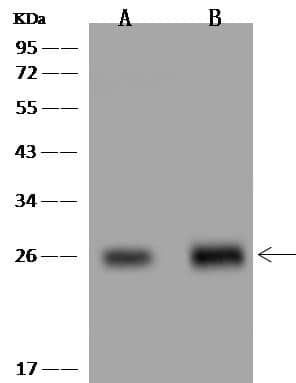
Western Blot-NBP3-20223-PDAP1 Antibody-Lane A: HeLa Whole Cell LysateLane B: U251 MG Whole Cell LysateLysates/proteins at 30 ug per lane.SecondaryGoat Anti-Rabbit IgG (H+L)/HRP at 1/10000 dilution.Developed using the technique.Performed under reducing conditions.Predicted band size:21 kDaObserved band size:26 kDaImmunoprecipitation-NBP3-20223-PDAP1 Antibody-
Immunoprecipitation-NBP3-20223-PDAP1 Antibody-Lane A:0.5 mg HeLa Whole Cell Lysate4 uL anti-PDAP1 rabbit polyclonal antibody and 60 ug of Immunomagnetic beads Protein A/G.Primary antibody:Anti-PDAP1 rabbit polyclonal antibody,at 1:100 dilutionSecondary antibody:Goat Anti-Rabbit IgG (H+L)/HRP at 1/10000 dilutionDeveloped using the ECL technique.Performed under reducing conditions.Predicted band size: 21 kDaObserved band size :26 kDaICC/IF-NBP3-20223-PDAP1 Antibody-
ICC/IF-NBP3-20223-PDAP1 Antibody-staining of PDAP1 in U251MG cells. Cells were fixed with 4% PFA,blocked with 10% serum, and incubated with rabbit anti-Human PDAP1 polyclonal antibody (dilution ratio 1:200) at 4C overnight. Then cells were stained with the Alexa Fluor®488-conjugated Goat Anti-rabbit IgG secondary antibody (green) and counterstained with DAPI (blue).Positive staining was localized to Cytoplasm and cell membrane.Applications for PDAP1 Antibody
Application
Recommended Usage
Immunocytochemistry/ Immunofluorescence
1:100-1:500
Immunoprecipitation
1-5 uL/mg lysate
Western Blot
1:500-1:2000
Formulation, Preparation, and Storage
Purification
Antigen and protein A Affinity-purified
Formulation
PBS
Preservative
0.03% Proclin 300
Concentration
Please see the vial label for concentration. If unlisted please contact technical services.
Shipping
The product is shipped with polar packs. Upon receipt, store it immediately at the temperature recommended below.
Stability & Storage
Store at 4C short term. Aliquot and store at -20C long term. Avoid freeze-thaw cycles.
Background: PDAP1
Alternate Names
HASPP28, PAP1PDGFA-associated protein 1, PAP28 kDa heat- and acid-stable phosphoprotein, PDGFA associated protein 1, PDGF-associated protein
Gene Symbol
PDAP1
Additional PDAP1 Products
Product Specific Notices for PDAP1 Antibody
This product is for research use only and is not approved for use in humans or in clinical diagnosis. Primary Antibodies are guaranteed for 1 year from date of receipt.
Loading...
Loading...
Loading...
Loading...


![Immunoprecipitation: PDAP1 Antibody [NBP3-20223] - PDAP1 Antibody](https://resources.bio-techne.com/images/products/nbp3-20223_rabbit-pdap1-pab-1262024149340.png)