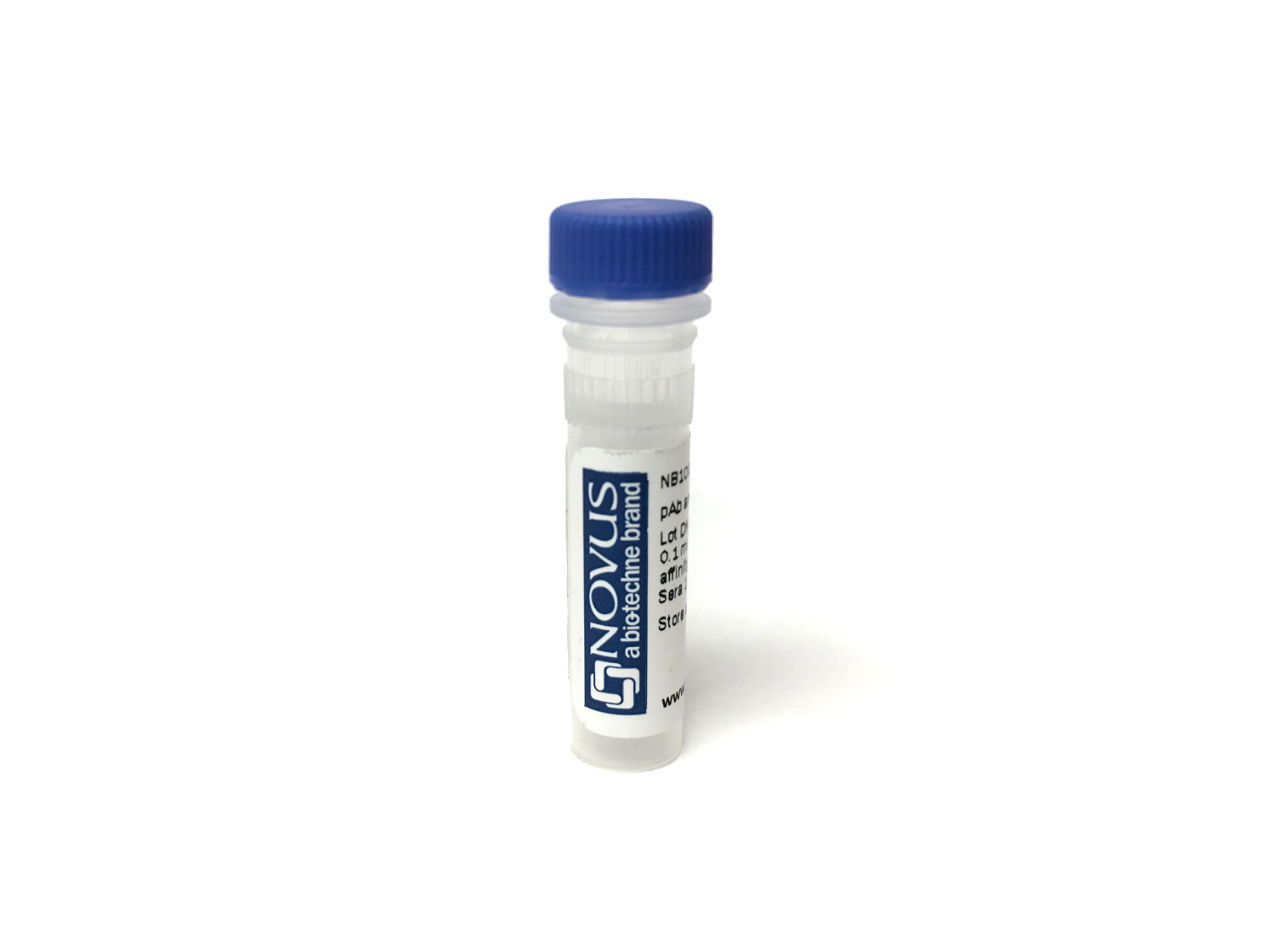
Proteinase 3/Myeloblastin/PRTN3 Antibody (PR3G-2)
Novus Biologicals, part of Bio-Techne | Catalog # NBP3-11271

Key Product Details
Species Reactivity
Human
Applications
Flow Cytometry, Functional, Immunoassay, Western Blot
Label
Unconjugated
Antibody Source
Monoclonal Mouse IgG1 Clone # PR3G-2
Concentration
0.1 mg/ml
Product Specifications
Immunogen
The monoclonal antibody PR3G-2 was produced by immunization of mice with a crude granule extract.
Clonality
Monoclonal
Host
Mouse
Isotype
IgG1
Endotoxin Level
<24 EU/mg
Applications for Proteinase 3/Myeloblastin/PRTN3 Antibody (PR3G-2)
Application
Recommended Usage
Flow Cytometry
Optimal dilutions of this antibody should be experimentally determined.
Functional
Optimal dilutions of this antibody should be experimentally determined.
Immunoassay
Optimal dilutions of this antibody should be experimentally determined.
Western Blot
Optimal dilutions of this antibody should be experimentally determined.
Application Notes
For Immunohistochemistry, Flow Cytometry and Western blotting dilutions to be used depend on detection system applied. It is recommended that users test the reagent and determine their own optimal dilutions. The typical starting working dilution is 1:50.
Formulation, Preparation, and Storage
Purification
Protein G purified
Formulation
0.2 um filtered solution in PBS, 0.1% BSA
Preservative
No Preservative
Concentration
0.1 mg/ml
Shipping
The product is shipped with polar packs. Upon receipt, store it immediately at the temperature recommended below.
Stability & Storage
Store at 4C.
Background: Proteinase 3/Myeloblastin/PRTN3
Alternate Names
ACPA, AGP7, C-ANCA, MBN, MBT, Myeloblastin, NP4, PRTN3
Gene Symbol
PRTN3
Additional Proteinase 3/Myeloblastin/PRTN3 Products
Product Documents for Proteinase 3/Myeloblastin/PRTN3 Antibody (PR3G-2)
Product Specific Notices for Proteinase 3/Myeloblastin/PRTN3 Antibody (PR3G-2)
This product is for research use only and is not approved for use in humans or in clinical diagnosis. Primary Antibodies are guaranteed for 1 year from date of receipt.
Loading...
Loading...
Loading...
Loading...
Loading...
