Rat CX3CL1/Fractalkine Chemokine Domain Antibody
R&D Systems, part of Bio-Techne | Catalog # AF537


Key Product Details
Species Reactivity
Validated:
Cited:
Applications
Validated:
Cited:
Label
Antibody Source
Product Specifications
Immunogen
Gln25-Gly100
Accession # O55145
Specificity
Clonality
Host
Isotype
Endotoxin Level
Scientific Data Images for Rat CX3CL1/Fractalkine Chemokine Domain Antibody
Chemotaxis Induced by CX3CL1/Fractalkine and Neutralization by Rat CX3CL1/Fractalkine Antibody.
Recombinant Rat CX3CL1/Fractalkine (Catalog # 536-FR) chemoattracts the BaF3 mouse pro-B cell line transfected with human CX3CR1 in a dose-dependent manner (orange line). The amount of cells that migrated through to the lower chemotaxis chamber was measured by Resazurin (Catalog # AR002). Chemotaxis elicited by Recombinant Rat CX3CL1/Fractalkine (40 ng/mL) is neutralized (green line) by increasing concentrations of Goat Anti-Rat CX3CL1/Fractalkine Chemokine Domain Antigen Affinity-purified Polyclonal Antibody (Catalog # AF537). The ND50 is typically 0.3-1.2 µg/mL.CX3CL1/Fractalkine in Mouse Brain.
CX3CL1/Fractalkine was detected in perfusion fixed frozen sections of mouse brain (cingulate cortex) using 1.7 µg/mL Goat Anti-Rat CX3CL1/Fractalkine Chemokine Domain Antigen Affinity-purified Polyclonal Antibody (Catalog # AF537) overnight at 4 °C. Tissue was stained with the Anti-Goat HRP-DAB Cell & Tissue Staining Kit (brown; Catalog # CTS008) and counterstained with hematoxylin (blue). View our protocol for Chromogenic IHC Staining of Frozen Tissue Sections.Detection of Rat CX3CL1/Fractalkine by Immunohistochemistry
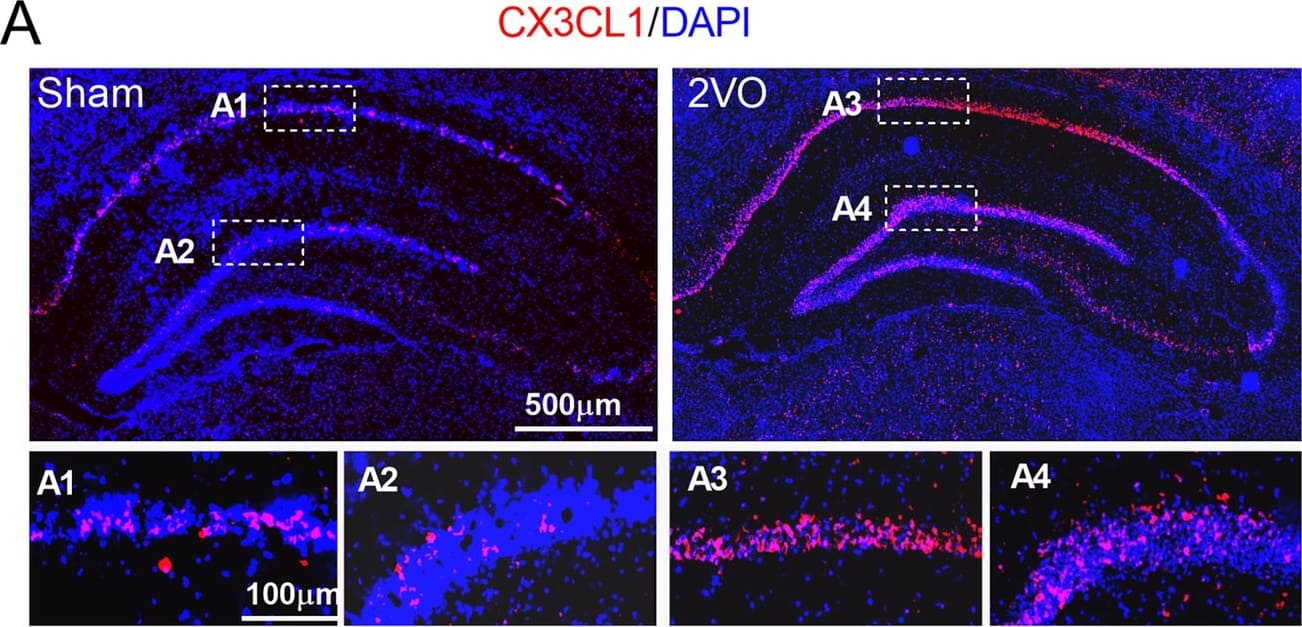
Upregulation of the expression of CX3CL1 and CX3CR1 in the hippocampus of 2VO and miR-195 loss-of-function rats. a, b Expression of CX3CL1 (red) and CX3CR1 (green) in the hippocampus of sham and 2VO rats was shown by immunofluorescence staining. The scale bar is 500 um. c Expression of CX3CL1 and CX3CR1 in the hippocampus of sham and 2VO rats detected by western blot technique. Bars represent the mean ± SD, n = 6, *P < 0.05 vs sham group, Student’s t test. d Lenti-pre-AMO-miR-195 upregulated the expression of CX3CL1 in rat hippocampus. Mean ± SD, n = 6, *P < 0.05 vs sham; #P < 0.05 vs lenti-pre-AMO-miR-195. Data were analyzed using one-way ANOVA followed by Tukey test. e Lenti-pre-AMO-miR-195 upregulated the expression of CX3CR1 in the rat hippocampus. Mean ± SD, n = 6, *P < 0.05 vs sham; #P < 0.05 vs lenti-pre-AMO-miR-195. Data were analyzed using one-way ANOVA followed by Tukey test Image collected and cropped by CiteAb from the following open publication (https://pubmed.ncbi.nlm.nih.gov/32819407), licensed under a CC-BY license. Not internally tested by R&D Systems.Applications for Rat CX3CL1/Fractalkine Chemokine Domain Antibody
Immunohistochemistry
Sample: Perfusion fixed frozen sections of mouse brain (cingulate cortex)
Western Blot
Sample: Recombinant Rat CX3CL1/Fractalkine (Catalog # 536-FR)
Neutralization
Rat CX3CL1/Fractalkine Sandwich Immunoassay
Reviewed Applications
Read 2 reviews rated 4 using AF537 in the following applications:
Formulation, Preparation, and Storage
Purification
Reconstitution
Formulation
Shipping
Stability & Storage
- 12 months from date of receipt, -20 to -70 °C as supplied.
- 1 month, 2 to 8 °C under sterile conditions after reconstitution.
- 6 months, -20 to -70 °C under sterile conditions after reconstitution.
Background: CX3CL1/Fractalkine
CX3CL1, also named neurotactin, is a member of the delta chemokine subfamily that contains a novel C-X3-C motif. Unlike other known chemokines, CX3CL1 is a type 1 membrane protein containing a chemokine domain tethered on a long mucin-like stalk. Rat CX3CL1 cDNA encodes a 393 amino acid (aa) residue precursor protein with two alternative (21 aa or 24 aa residue) putative signal peptides, a 74 aa or 76 aa residue globular chemokine domain, a 238 aa residue stalk region rich in Gly, Pro, Ser and Thr and containing degenerate mucin-like repeats, a 19 aa residue transmembrane segment and a 36 aa residue cytoplasmic domain. The extracellular domain of CX3CL1 can potentially be released as a soluble protein by proteolysis at the conserved dibasic motif proximal to the transmembrane region. With the exception of the stalk region, rat CX3CL1 shares a high degree of amino acid sequence homology (83% sequence identity) with human and mouse CX3CL1. CX3CL1 is expressed in various tissues including heart, brain, lung, kidney, skeletal muscle, and testis. In rat brain, CX3CL1 expression was found to be localized principally to neurons. The expression of CX3CL1 was also reported to be up-regulated on activated endothelial cells. Membrane-bound CX3CL1 has been shown to promote adhesion of leukocytes. The soluble chemokine domain of human CX3CL1 was reported to be chemotactic for T cells and monocytes while the soluble chemokine domain of mouse CX3CL1 was reported to chemoattract neutrophils and T‑lymphocytes but not monocytes. CX3CR1, previously named V28 or chemokine beta receptor‑like 1, has been found to be a specific receptor for CX3CL1. In addition, US28, a 7TM receptor encoded by human cytomegalovirus that binds multiple CC chemokines, has also been shown to bind CX3CL1 with high-affinity.
References
- Kledal, T.N. et al. (1998) FEBS Lett. 441:209.
- Combadiere, C. et al. (1998) J. Biol. Chem. 273:23799.
- Harrison, J.L. et al. (1998) Proc. Natl. Acad. Sci. USA 95:10896.
- Rossi, D.L. et al. (1998) Genomics 47:163.
Alternate Names
Gene Symbol
UniProt
Additional CX3CL1/Fractalkine Products
Product Documents for Rat CX3CL1/Fractalkine Chemokine Domain Antibody
Product Specific Notices for Rat CX3CL1/Fractalkine Chemokine Domain Antibody
For research use only

