Rat Fas/TNFRSF6/CD95 Antibody
R&D Systems, part of Bio-Techne | Catalog # AF2159


Key Product Details
Species Reactivity
Validated:
Cited:
Applications
Validated:
Cited:
Label
Antibody Source
Product Specifications
Immunogen
Gln22-Lys170
Accession # NP_631933
Specificity
Clonality
Host
Isotype
Scientific Data Images for Rat Fas/TNFRSF6/CD95 Antibody
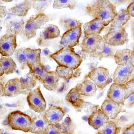
Fas/TNFRSF6/CD95 in Rat Liver.
Fas/TNFRSF6/CD95 was detected in perfusion fixed frozen sections of rat liver using Goat Anti-Rat Fas/TNFRSF6/CD95 Antigen Affinity-purified Polyclonal Antibody (Catalog # AF2159) at 15 µg/mL overnight at 4 °C. Tissue was stained using the Anti-Goat HRP-DAB Cell & Tissue Staining Kit (brown; Catalog # CTS008) and counterstained with hematoxylin (blue). Specific staining was localized to hepatocyte cell membranes. View our protocol for Chromogenic IHC Staining of Frozen Tissue Sections.Applications for Rat Fas/TNFRSF6/CD95 Antibody
CyTOF-ready
Flow Cytometry
Sample: Rat splenocytes
Immunohistochemistry
Sample: Perfusion fixed frozen sections of rat intestine, liver, and thymus
Western Blot
Sample: Recombinant Rat Fas/TNFRSF6/CD95 Fc Chimera (Catalog # 2159-FA)
Formulation, Preparation, and Storage
Purification
Reconstitution
Formulation
Shipping
Stability & Storage
- 12 months from date of receipt, -20 to -70 °C as supplied.
- 1 month, 2 to 8 °C under sterile conditions after reconstitution.
- 6 months, -20 to -70 °C under sterile conditions after reconstitution.
Background: Fas/TNFRSF6/CD95
Fas, also known as APO-1, CD95, and TNFRSF6, belongs to the death receptor family, which is a subfamily of the TNF receptor superfamily (1). Death receptors contain a cytoplasmic death domain (DD), which is required for transducing apoptotic signals. Engagement of Fas by its ligand (FasL) or agonistic anti-Fas antibodies induces dimerization and oligomerization of preformed Fas trimers. The activated receptor recruits the adaptor molecule FADD to form the Death-Inducing Signaling Complex (DISC) that also contains caspases. Upon activation, the caspases initiate a signaling cascade that induces the characteristic apoptotic phenotypes (2). Fas is highly expressed in epithelial cells, hepatocytes, activated mature lymphocytes, virus-transformed lymphocytes and other tumor cells. Fas expression has also been detected in mouse thymus, liver, heart, lung, kidney and ovary. FasL is a member of the TNF family of type 2 membrane proteins. FasL is predominantly expressed by activated T-lymphocytes, NK cells, and in tissues with immune-privileged sites (3).
Fas plays a role in the down-regulation of the immune reaction and has been shown to be an essential mediator of activation-induced death of activated T lymphocytes. Fas-mediated cell death has also been shown to be important for the deletion of activated or autoreactive B-lymphocytes. Both human and mice with genetic defects in Fas accumulate abnormal lymphocytes and develop systemic autoimmunity (4). Besides the perforin/granzyme-based mechanism, the Fas-FasL system has been identified as the alternate pathway for CTL-mediated cytotoxicity (5). FasL has also been shown to function in immunological privileged sites by killing infiltrating Fas-bearing lymphocytes and inflammatory cells (6). Rat Fas cDNA encodes a 324 amino acid residue type 1 membrane protein. The extracellular domain of rat Fas shares 54.1% and 66.7% amino acid sequence identity with that of human and mouse Fas, respectively.
References
- Ashkenazi, A. and V. Dixit (1999) Curr. Opin. Cell Biol. 11:255.
- Thorburn, S. (2003) Cellular Signaling 16:139.
- Green, D.R. and C.F. Ware (1997) Proc. Natl. Acad. Sci. USA 94:5986.
- Siegel, R.M. et al. (2003) Immunol. Res. 27:499.
- Barry, M. et al. (2000) Mol. Cell Biol. 20:3781.
- Barreiro, R. et al. (2004) J. Immunol. 173:1519.
Long Name
Alternate Names
Entrez Gene IDs
Gene Symbol
UniProt
Additional Fas/TNFRSF6/CD95 Products
Product Documents for Rat Fas/TNFRSF6/CD95 Antibody
Product Specific Notices for Rat Fas/TNFRSF6/CD95 Antibody
For research use only