Rat Neuropilin-2 Antibody
R&D Systems, part of Bio-Techne | Catalog # MAB567R


Key Product Details
Species Reactivity
Applications
Label
Antibody Source
Product Specifications
Immunogen
Gln23-Asp857 (Val809-Asp825 del)
Accession # O35276
Specificity
Clonality
Host
Isotype
Scientific Data Images for Rat Neuropilin-2 Antibody
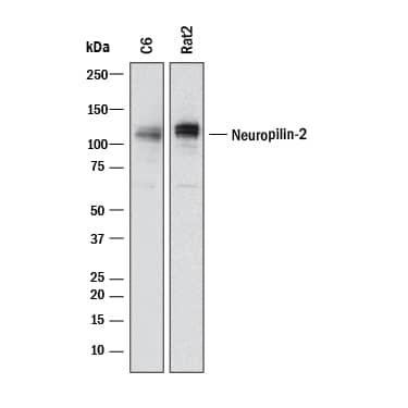
Detection of Rat Neuropilin‑2 by Western Blot.
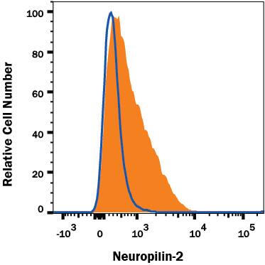
Western blot shows lysates of C6 rat glioma cell line and Rat-2 rat embryonic fibroblast cell line. PVDF membrane was probed with 1 µg/mL of Mouse Anti-Rat Neuropilin-2 Monoclonal Antibody (Catalog # MAB567R) followed by HRP-conjugated Anti-Mouse IgG Secondary Antibody (Catalog # HAF018). A specific band was detected for Neuropilin-2 at approximately 110 kDa (as indicated). This experiment was conducted under reducing conditions and using Immunoblot Buffer Group 1.Detection of Neuropilin-2 in C6 Rat Cell Line by Flow Cytometry.
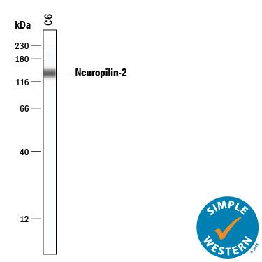
C6 Rat glial tumor cell line was stained with Mouse Anti-Rat Neuropilin-2 Monoclonal Antibody (Catalog # MAB567R, filled histogram) or isotype control antibody (Catalog # MAB0041, open histogram), followed by Allophycocyanin-conjugated Anti-Mouse IgG Secondary Antibody (Catalog # F0101B). View our protocol for Staining Membrane-associated Proteins.Detection of Rat Neuropilin‑2 by Simple WesternTM.
Simple Western lane view shows lysates of C6 rat glioma cell line, loaded at 0.2 mg/mL. A specific band was detected for Neuropilin‑2 at approximately 138 kDa (as indicated) using 50 µg/mL of Mouse Anti-Rat Neuropilin‑2 Monoclonal Antibody (Catalog # MAB567R) . This experiment was conducted under reducing conditions and using the 12-230 kDa separation system.Applications for Rat Neuropilin-2 Antibody
CyTOF-ready
Flow Cytometry
Sample: C6 Rat Cell Line
Simple Western
Sample: C6 rat glioma cell line
Western Blot
Sample: C6 rat glioma cell line and Rat‑2 rat embryonic fibroblast cell line
Formulation, Preparation, and Storage
Purification
Reconstitution
Formulation
Shipping
Stability & Storage
- 12 months from date of receipt, -20 to -70 °C as supplied.
- 1 month, 2 to 8 °C under sterile conditions after reconstitution.
- 6 months, -20 to -70 °C under sterile conditions after reconstitution.
Background: Neuropilin-2
Neuropilin-1 (Npn-1, previously known as neuropilin) and Npn-2 (previously known as Npn-1-related molecule) are type I transmembrane proteins that bind members of the class III secreted semaphorin subfamily which are implicated in repulsive axon guidance. The extracellular domain of these proteins is composed of two N-terminal CUB (complement-binding) domains (domains a1 and a2), two domains with homology to coagulation factors V and VIII (domains b1 and b2) and a MAM (meprin) domain (domain c). In the absence of ligands, neuropilins can form homo- and hetero-oligomers via homophilic interactions of their MAM domains. At the amino acid sequence level, Npn-1and Npn-2 share 44% identity. Npn-1 and Npn-2 show different binding specificities for different members of the semaphorin family. The expression patterns of Npn-1 and Npn-2 in developing neurons of the central and peripheral nervous systems are largely, though not completely non-overlapping. Npn‑1 and Npn-2 are also expressed by endothelial and tumor cells and have been shown to be isoform-specific receptors for VEGF165. Npn-1 was also reported to bind PlGF-2 and the VEGF-like protein from of virus NZ2.
References
- Fujisawa, H. and T. Kitsukawa (1998) Curr. Opin. Neurobiol. 8:587.
- Neufeld, G. et al. (1999) FASEB J. 13:9.
- Poltorak, Z. et al. (2000) J. Biol. Chem. 275:18040.
Alternate Names
Gene Symbol
UniProt
Additional Neuropilin-2 Products
Product Documents for Rat Neuropilin-2 Antibody
Product Specific Notices for Rat Neuropilin-2 Antibody
For research use only

