SIPA1 Antibody
Novus Biologicals, part of Bio-Techne | Catalog # NBP1-77947

Key Product Details
Species Reactivity
Human, Mouse
Applications
ELISA, Immunohistochemistry, Immunohistochemistry-Paraffin, Western Blot
Label
Unconjugated
Antibody Source
Polyclonal Rabbit IgG
Concentration
Please see the vial label for concentration. If unlisted please contact technical services.
Product Specifications
Immunogen
This affinity purified SIPA1 Antibody was prepared from whole rabbit serum produced by repeated immunizations with a synthetic peptide corresponding to a region near the amino terminus of mouse SIPA1. (Uniprot: P46062)
Reactivity Notes
A BLAST analysis was used to suggest cross-reactivity with SIPA1 from mouse, human and rat based on a 100% homology with the immunizing sequence. Cross-reactivity with SIPA1 from other sources has not been determined.
Localization
Perinuclear region
Clonality
Polyclonal
Host
Rabbit
Isotype
IgG
Description
This product was affinity purified from monospecific antiserum by immunoaffinity chromatography
Store vial at -20C prior to opening. Aliquot contents and freeze at -20C or below for extended storage. Avoid cycles of freezing and thawing. Centrifuge product if not completely clear after standing at room temperature. This product is stable for several weeks at 4C as an undiluted liquid. Dilute only prior to immediate use.
Store vial at -20C prior to opening. Aliquot contents and freeze at -20C or below for extended storage. Avoid cycles of freezing and thawing. Centrifuge product if not completely clear after standing at room temperature. This product is stable for several weeks at 4C as an undiluted liquid. Dilute only prior to immediate use.
Scientific Data Images for SIPA1 Antibody
Western Blot: SIPA1 Antibody [NBP1-77947]
Western Blot: SIPA1 Antibody [NBP1-77947] - Detection of over-expressed Sipa1 in lysates from mouse 3T3 cells transfected with Sipa1 (lane 1). Endogenous Sipa1 is detected in lane 2, which contains lysate from 3T3 cells mock-transfected with LacZGLB, although at a significantly reduced level compared to transfected cells. Lane 3 and 4 are similar to lanes 1 and 2 except the antibody was preincubated with the immunizing peptide prior to reaction with the membrane. The identity of the higher and lower molecular weight bands is unknown. The band at ~130 kDa, indicated by the arrowhead, corresponds to recombinant Sipa1. Primary antibody was used at 1:1250.Immunohistochemistry: SIPA1 Antibody [NBP1-77947]
Immunohistochemistry: SIPA1 Antibody [NBP1-77947] - Tissue: small intestine. Fixation: formalin fixed paraffin embedded. Primary antibody: Anti-Sipa1 at 5 ug/mL for 1 h at RT. Secondary antibody: Peroxidase rabbit secondary antibody at 1:10,000 for 45 min at RT. Staining: Sipa-1 as precipitated red signal with hematoxylin purple nuclear counterstain.Immunohistochemistry: SIPA1 Antibody [NBP1-77947] -
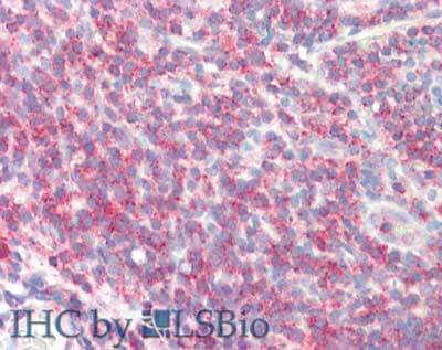
Immunohistochemistry: SIPA1 Antibody [NBP1-77947] - Affinity purified anti-Sipa1 antibody was used at 1.25 ug/ml to detect signal in a variety of tissues including multi-human, multi-brain and multi-cancer slides. This image shows moderate to strong positive staining of lymphocytes within human tonsil at 40X. Tissue was formalin-fixed and paraffin embedded. The image shows localization of the antibody as the precipitated red signal, with a hematoxylin purple nuclear counterstain. Personal Communication, Tina Roush, LifeSpanBiosciences, Seattle, WA.Applications for SIPA1 Antibody
Application
Recommended Usage
ELISA
1:20000
Immunohistochemistry
1.25-2.5 ug/ml
Immunohistochemistry-Paraffin
1:10-1:500
Western Blot
1:1000-1:5000
Application Notes
This affinity purified antibody has been tested for use in ELISA, immunohistochemistry, and western blotting. Specific conditions for reactivity should be optimized by the end user. Expect a band approximately 130 kDa in size corresponding
Formulation, Preparation, and Storage
Purification
Immunogen affinity purified
Formulation
0.02 M Potassium Phosphate, 0.15 M Sodium Chloride, pH 7.2
Preservative
0.01% Sodium Azide
Concentration
Please see the vial label for concentration. If unlisted please contact technical services.
Shipping
The product is shipped with polar packs. Upon receipt, store it immediately at the temperature recommended below.
Stability & Storage
Store at -20C short term. Aliquot and store at -80C long term. Avoid freeze-thaw cycles.
Background: SIPA1
Alternate Names
GTPase-activating protein Spa-1, MGC102688, MGC17037, p130 SPA-1, signal-induced proliferation-associated 1, signal-induced proliferation-associated protein 1, Sipa-1, SPA1signal-induced proliferation-associated gene 1
Gene Symbol
SIPA1
UniProt
Additional SIPA1 Products
Product Documents for SIPA1 Antibody
Product Specific Notices for SIPA1 Antibody
This product is for research use only and is not approved for use in humans or in clinical diagnosis. Primary Antibodies are guaranteed for 1 year from date of receipt.
Loading...
Loading...
Loading...
Loading...
![Immunohistochemistry: SIPA1 Antibody [NBP1-77947] Immunohistochemistry: SIPA1 Antibody [NBP1-77947]](https://resources.bio-techne.com/images/products/antibody/SIPA1-Antibody-Immunohistochemistry-NBP1-77947-img0005.jpg)
![Immunohistochemistry: SIPA1 Antibody [NBP1-77947] - SIPA1 Antibody](https://resources.bio-techne.com/images/products/nbp1-77947_rabbit-polyclonal-sipa1-antibody-235202318143119.jpg)

![Western Blot: SIPA1 Antibody [NBP1-77947] Western Blot: SIPA1 Antibody [NBP1-77947]](https://resources.bio-techne.com/images/products/antibody/SIPA1-Antibody-Western-Blot-NBP1-77947-img0002.jpg)