TSG101 Antibody - BSA Free
Novus Biologicals, part of Bio-Techne | Catalog # NBP2-77452

![Western Blot: TSG101 AntibodyBSA Free [NBP2-77452] Western Blot: TSG101 AntibodyBSA Free [NBP2-77452]](https://resources.bio-techne.com/images/products/TSG101-Antibody-Western-Blot-NBP2-77452-img0002.jpg)
Conjugate
Catalog #
Key Product Details
Species Reactivity
Human, Mouse, Rat
Applications
Immunocytochemistry/ Immunofluorescence, Immunohistochemistry, Immunohistochemistry-Paraffin, Western Blot
Label
Unconjugated
Antibody Source
Polyclonal Rabbit
Format
BSA Free
Concentration
1.0 mg/ml
Product Specifications
Immunogen
Partial synthetic peptide made to an internal portion of the human TSG101 protein (between amino acids 125-210) [UniProt Q99816]
Clonality
Polyclonal
Host
Rabbit
Scientific Data Images for TSG101 Antibody - BSA Free
Western Blot: TSG101 AntibodyBSA Free [NBP2-77452]
Western Blot: TSG101 Antibody [NBP2-77452] - Total protein from human HeLa, HepG2, mouse 3T3 and rat PC12 cells lines was separated on a 12% gel by SDS-PAGE, transferred to PVDF membrane and blocked in 5% non-fat milk in TBST. The membrane was probed with 2.0 ug/mL anti-TSG101 in blocking buffer and detected with an anti-rabbit HRP secondary antibody using West Pico PLUS chemiluminescence detection reagent.Immunocytochemistry/ Immunofluorescence: TSG101 Antibody - BSA Free [NBP2-77452]
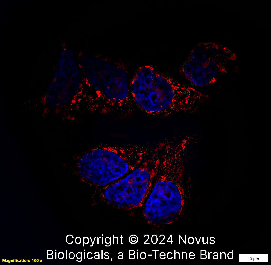
Immunocytochemistry/Immunofluorescence: TSG101 Antibody [NBP2-77452] - NIH3T3 cells were fixed for 10 minutes using 10% formalin and then permeabilized for 5 minutes using 1X PBS + 0.05% Triton X-100. The cells were incubated with anti-TSG101 at 2 ug/mL overnight at 4C and detected with an anti-rabbit DyLight 488 (Green) at a 1:500 dilution. Alpha tubulin (DM1A) NB100-690 was used as a co-stain at a 1:1000 dilution and detected with an anti-mouse DyLight 550 (Red) at a 1:500 dilution. Nuclei were counterstained with DAPI (Blue). Cells were imaged using a 40X objective.Immunohistochemistry-Paraffin: TSG101 Antibody - BSA Free [NBP2-77452]
Immunohistochemistry-Paraffin: TSG101 Antibody [NBP2-77452] - Analysis of a FFPE tissue section of the human small intestine using 1:200 dilution of TSG101 antibody (NBP2-77452). The signal was developed using HRP-DAB method which followed counterstaining of the cells with hematoxylin. The antibody generated staining mainly in the cytoplasm and membrane of glandular cells.Applications for TSG101 Antibody - BSA Free
Application
Recommended Usage
Immunocytochemistry/ Immunofluorescence
2 ug/ml
Immunohistochemistry
1:100 - 1:200
Immunohistochemistry-Paraffin
1:100 - 1:200
Western Blot
1 ug/ml
Formulation, Preparation, and Storage
Purification
Immunogen affinity purified
Formulation
PBS
Format
BSA Free
Preservative
0.02% Sodium Azide
Concentration
1.0 mg/ml
Shipping
The product is shipped with polar packs. Upon receipt, store it immediately at the temperature recommended below.
Stability & Storage
Store at 4C for up to 3 months. For longer storage, aliquot and store at -20C.
Background: TSG101
Upon its initial discovery, TSG101 was recognized as a tumor suppressor protein due to the identification of deletions within the TSG101 gene in human breast carcinomas. However, re-examination of the initial findings argued against this function and supported that TSG101 promotes tumorigenesis (2). In agreement with this role, TSG101 expression is upregulated in several types of cancer including breast, ovarian, and colorectal carcinoma.
References
1. Schmidt, O., & Teis, D. (2012). The ESCRT machinery. Current Biology. https://doi.org/10.1016/j.cub.2012.01.028
2. Jiang, Y., Ou, Y., & Cheng, X. (2013). Role of TSG101 in cancer. Frontiers in Bioscience. https://doi.org/10.2741/4099
3. Balut, C. M., Gao, Y., Murray, S. A., Thibodeau, P. H., & Devor, D. C. (2010). ESCRT-dependent targeting of plasma membrane localized KCa3.1 to the lysosomes. American Journal of Physiology - Cell Physiology. https://doi.org/10.1152/ajpcell.00120.2010
4. Su, V., & Lau, A. F. (2014). Connexins: Mechanisms regulating protein levels and intercellular communication. FEBS Letters. https://doi.org/10.1016/j.febslet.2014.01.013
Long Name
Tumor Susceptibility 101
Alternate Names
TSG10, VPS23
Gene Symbol
TSG101
Additional TSG101 Products
Product Documents for TSG101 Antibody - BSA Free
Product Specific Notices for TSG101 Antibody - BSA Free
This product is for research use only and is not approved for use in humans or in clinical diagnosis. Primary Antibodies are guaranteed for 1 year from date of receipt.
Loading...
Loading...
Loading...
Loading...
![Immunocytochemistry/ Immunofluorescence: TSG101 Antibody - BSA Free [NBP2-77452] Immunocytochemistry/ Immunofluorescence: TSG101 Antibody - BSA Free [NBP2-77452]](https://resources.bio-techne.com/images/products/TSG101-Antibody-Immunocytochemistry-Immunofluorescence-NBP2-77452-img0004.jpg)
![Immunohistochemistry-Paraffin: TSG101 Antibody - BSA Free [NBP2-77452] Immunohistochemistry-Paraffin: TSG101 Antibody - BSA Free [NBP2-77452]](https://resources.bio-techne.com/images/products/TSG101-Antibody-Immunohistochemistry-Paraffin-NBP2-77452-img0001.jpg)
![Immunocytochemistry/ Immunofluorescence: TSG101 Antibody - BSA Free [NBP2-77452] Immunocytochemistry/ Immunofluorescence: TSG101 Antibody - BSA Free [NBP2-77452]](https://resources.bio-techne.com/images/products/TSG101-Antibody-Immunocytochemistry-Immunofluorescence-NBP2-77452-img0003.jpg)
