Viral wnvNS3 Protease Antibody
R&D Systems, part of Bio-Techne | Catalog # MAB29074

Key Product Details
Species Reactivity
Applications
Label
Antibody Source
Product Specifications
Immunogen
sequences: rwnvNS2b 1-48, rwnvNS3 1-184
Specificity
Clonality
Host
Isotype
Scientific Data Images for Viral wnvNS3 Protease Antibody
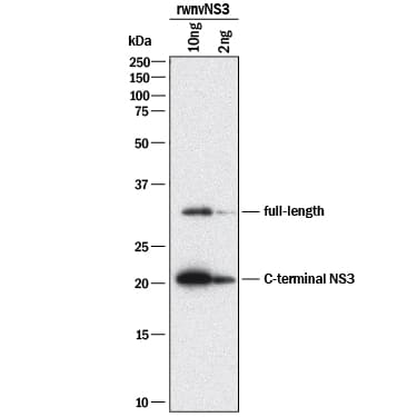
Detection of wnvNS3 Protease by Western Blot.
Western blot shows Recombinant Viral wnvNS3 Protease loaded at 10 µg and 2 µg. PVDF membrane was probed with 1 µg/mL of Mouse Anti-Viral wnvNS3 Protease Monoclonal Antibody (Catalog # MAB29074) followed by HRP-conjugated Anti-Mouse IgG Secondary Antibody (Catalog # HAF018). Specific bands were detected for wnvNS3 Protease at approximately 32 kDa for the full length and 22 kDa for the C-terminal NS3 (as indicated). This experiment was conducted under reducing conditions and using Immunoblot Buffer Group 1.Viral wnvNS3 Protease ELISA Standard Curve.
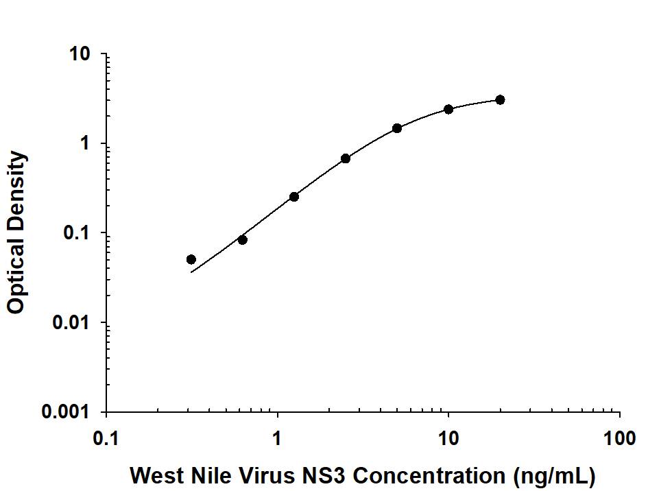
Recombinant Viral wnvNS3 Protease protein was serially diluted 2-fold and captured by Mouse Anti-Viral wnvNS3 Protease Monoclonal Antibody (Catalog # MAB29074) coated on a Clear Polystyrene Microplate (Catalog # DY990). Mouse Anti-Viral wnvNS3 Protease Monoclonal Antibody (Catalog # MAB29073) was biotinylated and incubated with the protein captured on the plate. Detection of the standard curve was achieved by incubating Streptavidin-HRP (Catalog # DY998) followed by Substrate Solution (Catalog # DY999) and stopping the enzymatic reaction with Stop Solution (Catalog # DY994).Applications for Viral wnvNS3 Protease Antibody
ELISA
This antibody functions as an ELISA capture antibody when paired with Mouse Anti-Viral wnvNS3 Protease Monoclonal Antibody (Catalog # MAB29073).
This product is intended for assay development on various assay platforms requiring antibody pairs.
Western Blot
Sample: Recombinant Viral wnvNS3 Protease
Formulation, Preparation, and Storage
Purification
Reconstitution
Formulation
Shipping
Stability & Storage
- 12 months from date of receipt, -20 to -70 °C as supplied.
- 1 month, 2 to 8 °C under sterile conditions after reconstitution.
- 6 months, -20 to -70 °C under sterile conditions after reconstitution.
Background: wnvNS3 Protease
Infection of mosquito-borne West Nile Virus can cause severe neurological disease and can be epidemic. Two non-structural proteins, NS3 and NS2b, play an essential role in viral replication and are therefore a potential target for treatment and prevention of West Nile Virus disease. NS3 consists of a trypsin-like serine protease with a catalytic triad (His51, Asp75, Ser135) and a putative helicase. Requiring NS2b as the cofactor, NS3 protease processes viral polyprotein precursor (1, 2). The purified recombinant protein used as an immunogen consists of three forms: the full-length fusion protein, the N-terminal NS2b, and the C-terminal NS3 with a G4SG4 linker. NS3 protease has a relatively narrow substrate specificity that prefers Arg in P1 and Lys in P2.
References
- Nall, T.A. et al. (2004) J. Biol. Chem. 279:48535.
- Chappell, K.J. et al. (2005) J. Biol. Chem. 274:2896.
Long Name
Entrez Gene IDs
Gene Symbol
Additional wnvNS3 Protease Products
Product Documents for Viral wnvNS3 Protease Antibody
Product Specific Notices for Viral wnvNS3 Protease Antibody
For research use only

