xCT Antibody - BSA Free
Novus Biologicals, part of Bio-Techne | Catalog # NB300-318


Conjugate
Catalog #
Key Product Details
Validated by
Orthogonal Validation, Biological Validation
Species Reactivity
Validated:
Human, Mouse, Rat
Cited:
Human, Mouse, Rat
Applications
Validated:
Dual RNAscope ISH-IHC, Flow Cytometry, Immunocytochemistry/ Immunofluorescence, Immunohistochemistry, Immunohistochemistry-Paraffin, Immunoprecipitation (Negative), Simple Western, Western Blot
Cited:
Block/Neutralize, Flow Cytometry, IF/IHC, Immunocytochemistry/ Immunofluorescence, Immunohistochemistry, Immunohistochemistry-Paraffin, In vivo assay, Western Blot
Label
Unconjugated
Antibody Source
Polyclonal Rabbit IgG
Format
BSA Free
Concentration
1.0 mg/ml
Product Specifications
Immunogen
This xCT Antibody was prepared from a synthetic peptide made to an N-terminal region of the human xCT protein (between residues 1-50) [UniProt Q9UPY5].
Reactivity Notes
Rat reactivity reported in scientific literature (PMID: 21540084).
Localization
Membrane
Clonality
Polyclonal
Host
Rabbit
Isotype
IgG
Scientific Data Images for xCT Antibody - BSA Free
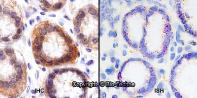
Dual RNAscope ISH-IHC: xCT Antibody [NB300-318] - Formalin-fixed paraffin-embedded tissue sections of human stomach were probed for xCT mRNA (ACD RNAScope Probe, catalog #422688; Fast Red chromogen, ACD catalog # 322750). Adjacent tissue section was processed for immunohistochemistry using Rabbit Polyclonal (Novus Biologicals catalog # NB300-318) at 0.25ug/mL with 1 hour incubation at room temperature followed by incubation with anti-rabbit IgG VisUCyte HRP Polymer Antibody (Catalog # VC003) and DAB chromogen (yellow-brown). Tissue was counterstained with hematoxylin (blue). Specific staining was localized to glandular cells.
Simple Western: xCT AntibodyBSA Free [NB300-318]
Simple Western: xCT Antibody [NB300-318] - Simple Western lane view shows a specific band for xCT using NB300-318 at 25 ug/ml in HeLa and HeLa + DEM cell lysates. This experiment was performed under reducing conditions using the 12-230 kDa separation system.Western Blot: xCT AntibodyBSA Free [NB300-318]
Western Blot: xCT Antibody [NB300-318] - Western blot analysis of protein isolated from subcutaneous tumors derived from in vivo growth of the clones relative to WT-derived tumors revealed that xCT levels remained low, phospho-STAT5 (p-STAT5) levels remained high, and phospho-STAT3 (p-STAT3) levels remained unchanged in the absence of SH-4-54. Image collected and cropped by CiteAb from the following publication (https://dx.plos.org/10.1371/journal.pone.0161202), licensed under a CC-BY license.Applications for xCT Antibody - BSA Free
Application
Recommended Usage
Flow Cytometry
2-3ug/ml. Use reported in scientific literature (PMID 20028852)
Immunocytochemistry/ Immunofluorescence
1:100-1:1000
Immunohistochemistry
5 u/gml
Immunohistochemistry-Paraffin
5 ug/ml
Simple Western
10 ug/ml
Western Blot
1:1000
Application Notes
Immunoprecipitation is not recommended. In Western blot this antibody recognizes a band at approx. 35 kDa, and in ICC/IF membrane staining was observed in HeLa cells. Permeablization is recommended prior to performing Flow analysis.
In Simple Western only 10 - 15 uL of the recommended dilution is used per data point. Separated by Size-Wes, Sally Sue/Peggy Sue.
In Simple Western only 10 - 15 uL of the recommended dilution is used per data point. Separated by Size-Wes, Sally Sue/Peggy Sue.
Reviewed Applications
Read 3 reviews rated 4.3 using NB300-318 in the following applications:
Formulation, Preparation, and Storage
Purification
Immunogen affinity purified
Formulation
PBS
Format
BSA Free
Preservative
0.02% Sodium Azide
Concentration
1.0 mg/ml
Shipping
The product is shipped with polar packs. Upon receipt, store it immediately at the temperature recommended below.
Stability & Storage
Store at 4C short term. Aliquot and store at -20C long term. Avoid freeze-thaw cycles.
Background: xCT/SLC7A11
References
1. Liu, J., Xia, X., & Huang, P. (2020). xCT: A Critical Molecule That Links Cancer Metabolism to Redox Signaling. Molecular therapy : the journal of the American Society of Gene Therapy. https://doi.org/10.1016/j.ymthe.2020.08.021
2. Koppula, P., Zhang, Y., Zhuang, L., & Gan, B. (2018). Amino acid transporter SLC7A11/xCT at the crossroads of regulating redox homeostasis and nutrient dependency of cancer. Cancer communications. https://doi.org/10.1186/s40880-018-0288-x
3. Lin, W., Wang, C., Liu, G., Bi, C., Wang, X., Zhou, Q., & Jin, H. (2020). SLC7A11/xCT in cancer: biological functions and therapeutic implications. American journal of cancer research.
4. xCT: Uniprot (Q9UPY5)
5. Koppula, P., Zhuang, L., & Gan, B. (2020). Cystine transporter SLC7A11/xCT in cancer: ferroptosis, nutrient dependency, and cancer therapy. Protein & cell. https://doi.org/10.1007/s13238-020-00789-5
6. Liu, L., Liu, R., Liu, Y., Li, G., Chen, Q., Liu, X., & Ma, S. (2020). Cystine-glutamate antiporter xCT as a therapeutic target for cancer. Cell biochemistry and function. https://doi.org/10.1002/cbf.3581
7. Cui, Q., Wang, J. Q., Assaraf, Y. G., Ren, L., Gupta, P., Wei, L., Ashby, C. R., Jr, Yang, D. H., & Chen, Z. S. (2018). Modulating ROS to overcome multidrug resistance in cancer. Drug resistance updates : reviews and commentaries in antimicrobial and anticancer chemotherapy. https://doi.org/10.1016/j.drup.2018.11.001
Long Name
Cationic 1/Solute Carrier Family 7 Member 11
Alternate Names
CCBR1, SLC7A11
Gene Symbol
SLC7A11
UniProt
Additional xCT/SLC7A11 Products
Product Documents for xCT Antibody - BSA Free
Product Specific Notices for xCT Antibody - BSA Free
This product is for research use only and is not approved for use in humans or in clinical diagnosis. Primary Antibodies are guaranteed for 1 year from date of receipt.
Loading...
Loading...
Loading...
Loading...
Loading...
Loading...
![Simple Western: xCT AntibodyBSA Free [NB300-318] Simple Western: xCT AntibodyBSA Free [NB300-318]](https://resources.bio-techne.com/images/products/xCT-Antibody-Simple-Western-NB300-318-img0013.jpg)
![Western Blot: xCT AntibodyBSA Free [NB300-318] Western Blot: xCT AntibodyBSA Free [NB300-318]](https://resources.bio-techne.com/images/products/xCT-Antibody-Western-Blot-NB300-318-img0015.jpg)
![Immunohistochemistry: xCT Antibody - BSA Free [NB300-318] Immunohistochemistry: xCT Antibody - BSA Free [NB300-318]](https://resources.bio-techne.com/images/products/xCT-Antibody-Immunohistochemistry-NB300-318-img0006.jpg)
![Flow Cytometry: xCT Antibody - BSA Free [NB300-318] Flow Cytometry: xCT Antibody - BSA Free [NB300-318]](https://resources.bio-techne.com/images/products/xCT-Antibody-Flow-Cytometry-NB300-318-img0009.jpg)
![Immunocytochemistry/ Immunofluorescence: xCT Antibody - BSA Free [NB300-318] Immunocytochemistry/ Immunofluorescence: xCT Antibody - BSA Free [NB300-318]](https://resources.bio-techne.com/images/products/xCT-Antibody-Immunocytochemistry-Immunofluorescence-NB300-318-img0005.jpg)
![Flow Cytometry: xCT Antibody - BSA Free [NB300-318] Flow Cytometry: xCT Antibody - BSA Free [NB300-318]](https://resources.bio-techne.com/images/products/xCT-Antibody-Flow-Cytometry-NB300-318-img0010.jpg)
![Western Blot: xCT AntibodyBSA Free [NB300-318] Western Blot: xCT AntibodyBSA Free [NB300-318]](https://resources.bio-techne.com/images/products/xCT-Antibody-Western-Blot-NB300-318-img0007.jpg)
![Flow Cytometry: xCT Antibody - BSA Free [NB300-318] Flow Cytometry: xCT Antibody - BSA Free [NB300-318]](https://resources.bio-techne.com/images/products/xCT-Antibody-Flow-Cytometry-NB300-318-img0008.jpg)
![Simple Western: xCT AntibodyBSA Free [NB300-318] Simple Western: xCT AntibodyBSA Free [NB300-318]](https://resources.bio-techne.com/images/products/xCT-Antibody-Simple-Western-NB300-318-img0014.jpg)
![Western Blot: xCT Antibody - BSA Free [NB300-318] - xCT Antibody - BSA Free](https://resources.bio-techne.com/images/products/nb300-318_rabbit-polyclonal-xct-antibody-310202415392520.jpg)
![Western Blot: xCT Antibody - BSA Free [NB300-318] - xCT Antibody - BSA Free](https://resources.bio-techne.com/images/products/nb300-318_rabbit-polyclonal-xct-antibody-31020241621818.jpg)
![Western Blot: xCT Antibody - BSA Free [NB300-318] - xCT Antibody - BSA Free](https://resources.bio-techne.com/images/products/nb300-318_rabbit-polyclonal-xct-antibody-310202416212337.jpg)