Mammary Epithelial Cell Growth Media
R&D Systems, part of Bio-Techne | Catalog # CCM028
View Terms and Conditions

Key Product Details
.")
Summary for Mammary Epithelial Cell Growth Media
Media for the serum-free expansion of mammary epithelial cells.
Key Benefits
- Unique and optimized formulation that promotes mammary epithelial cell growth
- Includes a single supplement featuring key growth factors and small molecules
- 250 mL bottle results in less wasted media
- Quality control tested for consistent media performance

Why Use Pre-optimized, Serum-free Media to Grow Endothelial Cells?
Primary mammary epithelial cells are commonly used for the study of disease mechanisms and drug discovery in breast tissue cancer/disease. Mammary epithelial cells are phenotypically characterized as Cytokeratin 14+, Cytokeratin 18+, and Cytokeratin 19-.
R&D Systems® Mammary Epithelial Cell Growth Media is formulated to support the expansion of human primary mammary epithelial cells under serum-free conditions. The media has been tested for its ability to support Cytokeratin 14+, Cytokeratin 18+, and Cytokeratin 19- human mammary epithelial cell growth in vitro.
This kit contains the following reagents to maintain mouse pluripotent stem cells in culture:
- Mammary Epithelial Cell Growth Base Media (250 mL)
- Mammary Epithelial Cell Growth Supplement (50X)
Note: The components for this kit require different storage/shipping temperatures and will arrive in separate packaging.
Stability and Storage
Store in the dark at ≤ -20 °C in a manual defrost freezer. Do not use past the expiration date.
Precaution
When handling biohazardous materials such as human cells, safe laboratory procedures should be followed and protective clothing should be worn.
Limitations
- FOR LABORATORY RESEARCH USE ONLY. NOT FOR USE IN DIAGNOSTIC PROCEDURES.
- The safety and efficacy of this product in diagnostic or other clinical uses has not been established.
- This reagent should not be used beyond the expiration date indicated on the label.
- Results may vary due to variations among primary epithelial cells originating from different donors.
") | Mammary Epithelial Cell Growth Media Supports Cell Proliferation of Human Mammary Epithelial Cells (HMECs). One well of a 6-well plate of HMECs (6.7 x 10⁴) cells was expanded over 5 passages using Complete Mammary Epithelial Cell Growth Media. Cells were passaged every 3-5 days. The histogram shows the cumulative number of HMECs expanded over 5 passages if all cells were replated at each passage. |
 Express Mammary Epithelial Cell Markers Cytokeratin 14, Cytokeratin 18, and E-Cadherin") | Human Mammary Epithelial Cells (HMECs) Express Mammary Epithelial Cell Markers Cytokeratin 14, Cytokeratin 18, and E-Cadherin. HMECs were expanded in Complete Mammary Epithelial Cell Growth Media for 5 passages prior to staining for marker expression by immunocytochemistry. Cells were double stained with Goat Anti-Mouse E-Cadherin (green; R&D Systems®, Catalog # AF648) and either (A) Mouse Anti-Human Cytokeratin 14 Antibody (red; R&D Systems®, Catalog # MAB3164) or (B) Mouse Anti-Human Cytokeratin 18 Antibody (red; R&D Systems®, Catalog # 5748). The nuclei were counterstained with DAPI (blue; Tocris®, Catalog # 5748). |
|
|
Mammary Epithelial Cell Growth Media Supports Cell Proliferation of Human Mammary Epithelial Cells (HMECs). One well of a 6-well plate of HMECs (6.7 x 10⁴) cells was expanded over 5 passages using Complete Mammary Epithelial Cell Growth Media. Cells were passaged every 3-5 days. The histogram shows the cumulative number of HMECs expanded over 5 passages if all cells were replated at each passage. |
 Express Mammary Epithelial Cell Markers Cytokeratin 14, Cytokeratin 18, and E-Cadherin.")
|
Human Mammary Epithelial Cells (HMECs) Express Mammary Epithelial Cell Markers Cytokeratin 14, Cytokeratin 18, and E-Cadherin. HMECs were expanded in Complete Mammary Epithelial Cell Growth Media for 5 passages prior to staining for marker expression by immunocytochemistry. Cells were double stained with Goat Anti-Mouse E-Cadherin (green; R&D Systems®, Catalog # AF648) and either (A) Mouse Anti-Human Cytokeratin 14 Antibody (red; R&D Systems®, Catalog # MAB3164) or (B) Mouse Anti-Human Cytokeratin 18 Antibody (red; R&D Systems®, Catalog # 5748). The nuclei were counterstained with DAPI (blue; Tocris®, Catalog # 5748). |
Assay Procedure
Refer to the product datasheet for complete product details.
Briefly, Complete Mammary Epithelial Cell Growth Media is prepared using the following procedure:
- Add Mammary Epithelial Cell Growth Media Supplement to Mammary Epithelial Cell Growth Base Media
- Store completed media at 2 – 8 °C for up to 2 weeks

Reagents provided with the Mammary Epithelial Cell Growth Media (Catalog # CCM028):
- Mammary Epithelial Cell Growth Base Media (250 mL)
- Mammary Epithelial Cell Growth Supplement (50X)
Note: The components for this kit require different storage/shipping temperatures and will arrive in separate packaging.
Reagents
- Human mammary epithelial cells (HMECs)
- Penicillin-Streptomycin (100X), optional
- TrypLE™ Express
- Phosphate-Buffered Saline (PBS) (Tocris; Catalog # 3156)
Materials
- 10 cm tissue culture dishes
- 15 mL centrifuge tubes
- Serological pipettes
- Pipette and pipette tips
Equipment
- 37 °C and 5% CO2 incubator
- Centrifuge (low speed clinical or equivalent)
- Hemocytometer
- Inverted Microscope
- Water bath
Reagent Preparation
Mammary Epithelial Cell Growth Supplement - Thaw the Mammary Epithelial Cell Growth Supplement at 2-8 °C or room temperature.
Complete Mammary Epithelial Cell Growth Media - Add 5 mL of Mammary Epithelial Cell Growth Supplement to 250 mL of Mammary Epithelial Cell Growth Base Media. Add Penicillin-Streptomycin (100X) at a 1:100 dilution. Store under sterile conditions at 2-8 °C for up to 2 weeks. Note: If Penicillin-Streptomycin is not needed for the experiment, it can be omitted.
Culturing of HMECs
Warm Complete Mammary Epithelial Cell Growth Media to 37 °C.
Determine the size and number of dishes needed for plating.

Warm the frozen vial of HMECs until just thawed.

Transfer cells immediately and gently into a 15 mL centrifuge tube containing > 5 mL of prewarmed Complete Mammary Epithelial Cell Growth Media.
Centrifuge at 200 x g for 5 minutes.
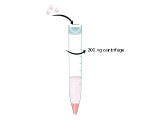
Resuspend the pellet in prewarmed Complete Mammary Epithelial Cell Growth Media.
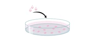
Transfer Transfer HMEC suspension to the dish in a total of 10 mL/10 cm dish.
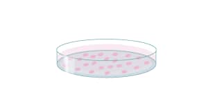
Change media the day following thaw.
Monitor cells daily and change media every other day.
Passage the cells at 80-90 % confluency.
Subculturing of HMECs
Warm Mammary Epithelial Cell Growth Media to 37 °C.
Remove and discard the media from the tissue culture dish.
Rinse cells once with sterile 1X PBS.
Digest cells with TrypLE™ Express for 3-5 minutes at 37 °C.
Tap the side of the dish to aid the detachment of the cells.
Transfer the cell suspension to a 15 mL centrifuge tube containing 8 mL of prewarmed Mammary Epithelial Cell Growth Media.
Centrifuge at 200 x g for 5 minutes.
Plate cells (approximately 6.7 x 10³ cells/cm2).
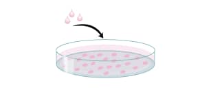
Change media the day following thaw.
Monitor cells daily and change media every other day.
Passage the cells at 80-90 % confluency.