Breadcrumb
- Home
- Products
- Hematopoietic Stem Cells
- Hematopoietic Stem Cells Cell Culture Products
- StemXVivo Serum-Free Human T Cell Base Media (CCM010)
StemXVivo Serum-Free Human T Cell Base Media
R&D Systems, part of Bio-Techne | Catalog # CCM010

Key Product Details

Summary for StemXVivo Serum-Free Human T Cell Base Media
Pre-optimized for ex vivo culture of human T lymphocytes.
Key Benefits
- Supports T lymphocyte expansion as well as or better than RPMI containing FBS
- Defined media decreases experimental variation
- High lot-to-lot consistency decreases variation
Why use pre-optimized media for ex vivo culture of human T lymphocytes?
Isolation of human T-lymphocytes from peripheral blood is a time consuming process that can yield cells of varying purity and quality.
Once in culture, the maintenance and expansion of T lymphocytes is highly dependent on the media, growth factors, and cytokines. R&D Systems offers Human StemXVivo® Serum-Free T Cell Base Media, a defined media that outperforms RPMI containing Fetal Bovine Serum in its ability to support T cell expansion.
Human StemXVivo® Serum-Free T Cell Base Media:
- Supports T lymphocyte expansion better than RPMI containing 10% FBS.
- Decreases experimental variability through the use of defined media components.
- Can be used for multiple applications including mixed lymphocyte reactions (MLR), expansion of antigen-specific or non-specific T cells, and proliferation of peripheral blood lymphocytes in the presence of pharmacological agents such as PMA or ConA
- 250 mL StemXVivo® Serum-Free T Cell Base Media.
The quantity of each component in this kit is sufficient to differentiate two 24-well plates, or an equivalent surface area, of pluripotent stem cells into hepatocyte-like cells
Stability and Storage
Upon receipt, the Human StemXVivo® Serum-Free T Cell Base Media should be stored at =-20° C in a manual defrost freezer. The media can be thawed at 2 °C to 8 °C or at room temperature. Thawed media can be aliquoted and stored at =-20 °C in a manual defrost freezer until the expiration date on the vial label or used within a month when stored in the dark at 2 °C to 8 °C. Avoid repeated freeze-thaw cycles.
Precautions
The human origin-derived components used in this product have been derived from human plasma, which has been tested and found negative for antibodies to HIV-1/2, hepatitis B surface antigen (HBsAg), and hepatitis C virus (HCV). However, the medium should be handled as if potentially infectious. Safe laboratory procedures should be followed and protective clothing should be worn when handling this media. The acute and chronic effects of over-exposure to this media are unknown.
Limitations
- FOR LABORATORY RESEARCH USE ONLY. NOT FOR USE IN DIAGNOSTIC PROCEDURES.
- The safety and efficacy of this product in diagnostic or other clinical uses has not been established.
- This reagent should not be used beyond the expiration date indicated on the label.
- Results may vary due to variations among primary T lymphocyte populations derived from different donors.
| HSC Differentiation &Lineage Marker Schematic View Interactive Pathway |
The definitive hematopoietic system is made up of all adult blood cell types including megakaryocytes, erythrocytes, and cells of the myeloid and lymphoid lineages. All of these cells are derived from multipotent hematopoietic stem cells (HSCs) through a succession of precursors with progressively limited potential. Hematopoietic stem cells are tissue-specific stem cells that exhibit remarkable self-renewal capacity and are responsible for the life-long maintenance of the hematopoietic system. HSCs are rare cells that reside in adult bone marrow where hematopoiesis is continuously taking place. They can also be found in cord blood, fetal liver, adult spleen, and peripheral blood. R&D Systems offers several products for studying hematopoietic lineage cells including serum-free media, lineage depletion antibodies and kits, and reagents for performing colony forming cell (CFC) assays.
| Species | Human |
| Source | N/A |

|
Proliferative Response of Cultured CD3/CD28 Primed T cells in Human StemXVivo® Serum-Free T Cell Base Media. 1 x 10⁵purified CD3+T cells were cultured for five days in StemXVivo®Serum-Free T Cell Base Media (Catalog # CCM010) or with RPMI supplemented with 10% Fetal Bovine Serum (RPMI/10% FBS). The purified CD3+T cells were cultured on 96 well microplates coated with Mouse Anti-Human CD3 epsilon (Clone UCHT1; Catalog # MAB100) and Goat Anti-Human CD28 Antigen Affinity-purified Polyclonal Antibody. [3H]-thymidine was added for the final 18 hours. Cells were harvested and the incorporation of [3H]-thymidine was measured using a beta-scintillation counter. Results are presented as the mean ± standard deviation of samples run in triplicate. |

|
Phenotypic Analysis of Cultured Monocyte-derived Dendritic Cells Before and After LPS-induced Maturation. Immature monocyte-derived dendritic cells (MoDCs) (open histograms; green line) were obtained after CD14+-enriched monocytes were cultured for seven days in StemXVivo®Serum-Free Dendritic Cell Base Media (Catalog # CCM003) supplemented with 50 ng/mL Recombinant Human GM-CSF (Catalog # 215-GM) and 35 ng/mL Recombinant Human IL-4 (Catalog # 204-IL). Mature monocyte-derived dendritic cells (filled histograms) were cultured under the same conditions for seven days and then induced with LPS for an additional 48 hours. Day seven immature MoDCs and day nine LPS-treated MoDCs were stained with a FITC-conjugated Mouse Anti-Human B7-1/CD80 Monoclonal Antibody (Catalog # FAB140F), a FITC-conjugated Mouse Anti-Human CD83 Monoclonal Antibody (Catalog # FAB1774F), a FITC-conjugated Mouse Anti-Human B7-2/CD86 Monoclonal Antibody (Catalog # FAB141F), an Anti-MHC Class II Antibody, or an appropriate isotype control antibody (empty histogram with solid line). |

|
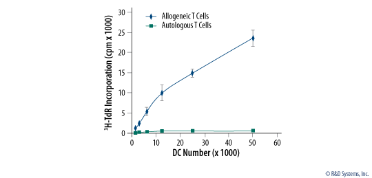
Mature Monocyte-derived Dendritic Cells Induce Proliferation of Allogenic T Cells CD14+monocytes were cultured for seven days in Human StemXVivo®Serum-free Dendritic Cell Base Media (Catalog # CCM003) supplemented with Recombinant Human GM-CSF (Catalog # 215-GM), Recombinant Human IL-4 (Catalog # 204-IL) and Gentamycin. The cells were subsequently treated with LPS for an additional 48 hours to induce dendritic Cell Differentiation/Maturation. Graded doses of mature monocyte-derived dendritic cells were incubated with 1 x 10⁵autologous or allogenic CD3+T cells for five days.3H-thymidine (3H-TdR) was added to the culture for the final 18 hours and T cell proliferation was measured using a scintillation counter. Results are presented as the mean cpm obtained from three experiments. |
Preparation & Storage
| Shipping Conditions | The product is shipped with dry ice or equivalent. Upon receipt, store it immediately at the temperature recommended below. |
| Storage | Store the unopened product at -20 to -70 °C. Use a manual defrost freezer and avoid repeated freeze-thaw cycles. Do not use past expiration date. |
Assay Procedure
Refer to the product datasheet for complete product details.
CD3/CD28-primed T lymphocytes can be cultured ex vivo in StemXVivo® Serum-Free T Cell Base Media using the following procedure:
- Coat a 96-well microplate with CD3 and CD28 antibodies
- Resuspend CD3+ T cells in StemXVivo® Serum-Free T Cell Base Media
- Add CD3+ T cells to the microplate
- Assess proliferation after 3-5 days
View Graphic Procedure
Reagent Supplied in StemXVivo® Serum-Free T Cell Base Media (Catalog # CCM010):
- 250 mL StemXVivo® Serum-Free T Cell Base Media
Reagents
- CD3+ lymphocytes isolated from PBMCs using MagCellect Human CD3+ T Cell Isolation Kit (Catalog # MAGH101), or Cell Enrichment Column (Catalog # HTCC)
- Human CD3 monoclonal antibody (Catalog # MAB100)
- Human CD28 polyclonal antibody (Catalog # AF-342-PB)
- Penicillin-Streptomycin
- Recombinant Human IL-2 (Catalog # 202-IL, optional)
- [3H]-thymidine
Materials
- 96 well v- or round-bottom microplate
- 15 mL centrifuge tubes
- Serological pipettes
- Pipettes and pipette tips
- Hemocytometer
- Cell harvester
Equipment
- 37 °C and 5% CO2 incubator
- Centrifuge
- Vortex Mixer
- Inverted microscope
- Beta-scintillation counter
Protocol for Culturing CD3/CD28-Primed T Lymphocytes Ex Vivo using StemXVivo® Serum-free T Cell Base Media (Catalog # CCM010)
Preparation of the CD3/CD28 coated 96-well microplate
- Add working solution of CD3 and CD28 antibody to each well of a 96-well microplate.
- Incubate for 90 minutes at 37 °C or at 2 °C to 8 °C overnight.
- Wash the wells with PBS three times
Thawing StemXVivo® Serum-Free T Cell Base Media and Plating CD3+ T Cells
- Thaw the Human StemXVivo® Serum-Free T Cell Base Media
- Prepare CD3+ T cells from peripheral blood mononuclear cells.
- Resuspend the purified CD3+ T cells in Human StemXVivo® Serum-Free T Cell Base Media.
- Perform a cell count and adjust the cell density to 1 x 10⁶ cells/mL.
- Add the CD3+ T cells to each well at 2 x 10⁵ cells in 0.2 mL.
- Place the plate in a humidified 37° C and 5% CO2 incubator for 3-5 days for short-term stimulation.
- Assess the proliferation of CD3/CD28-primed T cells by adding [3H]-thymidine to each well during the last 18 hours of a 3-5 day period.
- Harvest cells using a cell harvester and measure the cpm (counts per minute) with a beta-scintillation counter.