Mouse IgG1 Isotype Control Best Seller
R&D Systems, part of Bio-Techne | Catalog # MAB002


Key Product Details
Species Reactivity
Applications
Label
Antibody Source
Product Specifications
Immunogen
Specificity
Clonality
Host
Isotype
Endotoxin Level
Scientific Data Images for Mouse IgG1 Isotype Control
Detection of Mouse IgG1Isotype Control by Flow Cytometry
Human peripheral blood mononuclear cells (PBMCs) were stained with Mouse Anti-Human LILRA6/CD85b Monoclonal Antibody (Catalog # MAB8656, filled histogram) or Mouse IgG1Isotype Control Antibody (Catalog # MAB002, open histogram), followed by Allophycocyanin-conjugated Anti-Mouse IgG Secondary Antibody (Catalog # F0101B).Detection of Human IgG1 by Flow Cytometry
EC and PC ICAM-1 expression and effects of CD18 blocking on neutrophil adhesion.ICAM-1 expression on (A) EC or (D) PC monolayers before and after 4 hr IL-1 beta activation was assessed by flow cytometry (IgG labeled control white). ICAM-1 expression on (B,C) EC and (E,F) PC following 4 hr IL-1 beta activation was imaged using confocal microscopy. Scale bars are 20 µm. (G) Neutrophil adhesion inhibition to IL-1 beta activated EC or PC monolayers. Freshly isolated neutrophils were pre-incubated with anti-Mac-1 or anti-LFA-1 antibodies and seeded onto EC or PC monolayers (activated for 4 with IL-1 beta) in Sykes-Moore chambers and allowed to adhere prior to counting. Bars represent average neutrophil adhesion ± SEM. *P<0.05, when compared to the no block control. Image collected and cropped by CiteAb from the following publication (https://dx.plos.org/10.1371/journal.pone.0060025), licensed under a CC-BY license. Not internally tested by R&D Systems.Detection of Human IgG1 by Flow Cytometry
EC and PC ICAM-1 expression and effects of CD18 blocking on neutrophil adhesion.ICAM-1 expression on (A) EC or (D) PC monolayers before and after 4 hr IL-1 beta activation was assessed by flow cytometry (IgG labeled control white). ICAM-1 expression on (B,C) EC and (E,F) PC following 4 hr IL-1 beta activation was imaged using confocal microscopy. Scale bars are 20 µm. (G) Neutrophil adhesion inhibition to IL-1 beta activated EC or PC monolayers. Freshly isolated neutrophils were pre-incubated with anti-Mac-1 or anti-LFA-1 antibodies and seeded onto EC or PC monolayers (activated for 4 with IL-1 beta) in Sykes-Moore chambers and allowed to adhere prior to counting. Bars represent average neutrophil adhesion ± SEM. *P<0.05, when compared to the no block control. Image collected and cropped by CiteAb from the following publication (https://dx.plos.org/10.1371/journal.pone.0060025), licensed under a CC-BY license. Not internally tested by R&D Systems.Applications for Mouse IgG1 Isotype Control
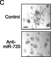
Control
Reviewed Applications
Read 22 reviews rated 4.6 using MAB002 in the following applications:
Formulation, Preparation, and Storage
Purification
Reconstitution
Formulation
Shipping
Stability & Storage
- 12 months from date of receipt, -20 to -70 °C as supplied.
- 1 month, 2 to 8 °C under sterile conditions after reconstitution.
- 6 months, -20 to -70 °C under sterile conditions after reconstitution.
Background: IgG1
R&D Systems offers a range of secondary antibodies and controls for flow cytometry, immunohistochemistry, and Western blotting. We provide species-specific secondary antibodies that are available with a variety of conjugated labels.
Our NorthernLights fluorescent secondary antibodies are bright and resistant to photobleaching. We are currently offering secondary antibodies recognizing mouse, rat, goat, sheep, and rabbit IgG as well as chicken IgY. These reagents are available with three distinct excitation and emission maxima, making them ideal for multi-color fluorescence microscopy.
Long Name
Alternate Names
Additional IgG1 Products
Product Documents for Mouse IgG1 Isotype Control
Product Specific Notices for Mouse IgG1 Isotype Control
For research use only


