Bovine Fibronectin Protein, CF Best Seller
R&D Systems, part of Bio-Techne | Catalog # 1030-FN

Key Product Details
Source
Conjugate
Applications
Product Specifications
Source
Purity
Endotoxin Level
Activity
In this application, the recommended concentration for this effect is
1-5 µg/cm2. Fibronectin can also be added in the media to support cell spreading at a concentration of 0.5-50 µg/mL.
Optimal concentration depends on cell type as well as the application or research objectives.
Measured by the ability of the immobilized protein to support the adhesion of B16-F1 mouse melanoma cells.
The ED50 for this effect is 10.0-100 ng/mL.
Reviewed Applications
Read 1 review rated 4 using 1030-FN in the following applications:
Scientific Data Images for Bovine Fibronectin Protein, CF
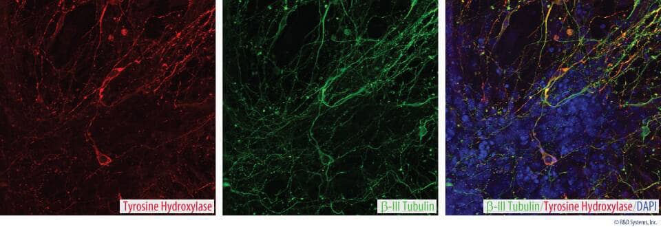
Culture and Characterization of Dopaminergic Neurons Generated from Human Pluripotent Stem Cells.
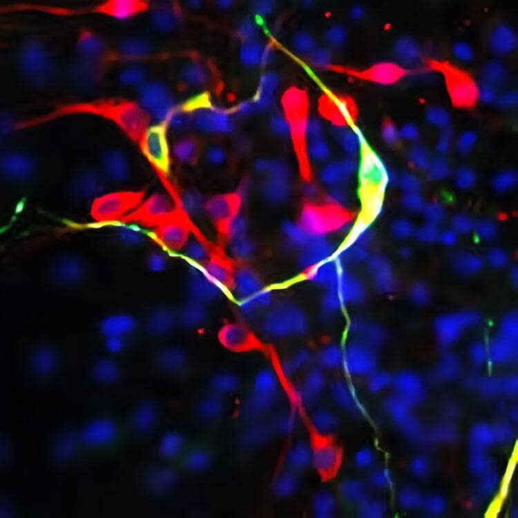
Dopaminergic neurons were generated from human pluripotent stem cells in media that included Bovine Fibronectin Protein (Catalog # 1030-FN) to support cell attachment and spreading, the ITS and N-2 Plus Media Supplements (AR013 and AR003, respectively) to select and enrich for neural stem cell populations, and a panel of growth factors for effective dopaminergic differentiation, including Recombinant Human FGF-basic, Recombinant Mouse FGF-8b (423-F8), and Recombinant Mouse Shh-N (464-SH). Cells were stained with a Mouse Anti-Human/Mouse Tyrosine Hydroxylase Monoclonal Antibody (MAB7566) followed by a NorthernLights™ 493-conjugated Donkey Anti-Mouse IgG Antigen Affinity-purified Secondary Antibody (NL009; green), and a Mouse Neuron-specific beta III Tubulin Tuj1 Monoclonal Antibody (MAB1195) followed by a NorthernLights 557-conjugated Donkey Anti-Mouse IgG Antigen Affinity-purified Secondary Antibody (NL007; red) and counterstained with DAPI (5748; blue).Culture and Characterization of Dopaminergic Neurons Generated from Human Pluripotent Stem Cells.
Dopaminergic neurons were generated from human pluripotent stem cells in media that included Bovine Fibronectin Protein (Catalog # 1030-FN) to support cell attachment and spreading, the ITS and N-2 Plus Media Supplements (AR013 and AR003, respectively), and a panel of growth factors for effective dopaminergic differentiation, including Recombinant Human FGF-basic, Recombinant Mouse FGF-8b (423-F8), and Recombinant Mouse Shh-N (464-SH). Tyrosine Hydroxylase was detected using a Mouse Anti-Human Tyrosine Hydroxylase Monoclonal Antibody (MAB7566). The cells were stained with the NorthernLights™ 557-conjugated Donkey Anti-Mouse IgG Antigen Affinity-purified Secondary Antibody (NL007; red). Neuron-specific beta-III Tubulin was detected using a Mouse Anti-Neuron-specific beta-III Tubulin (Clone Tuj-1) Monoclonal Antibody (MAB1195) followed by the NorthernLights™ 493-conjugated Donkey Anti-Mouse IgG Antigen Affinity-purified Secondary Antibody (NL009; green). Cells were counterstained with DAPI (5748; blue).Culture and Characterization of Mouse Oligodendrocytes.
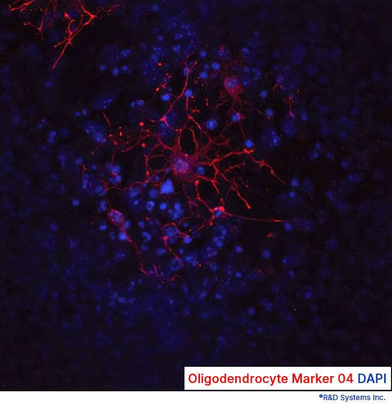
D3 mouse embryonic stem cells were expanded in KO-ES Media supplemented with Bovine Fibronectin Protein (Catalog # 1030-FN) to support cell attachment and spreading, the ITS and N-2 Plus Media Supplements (AR013 and AR003), and a panel of growth factors for effective oligodendrocyte differentiation, including Recombinant Human FGF-basic, Recombinant Human EGF (236-EG), and Recombinant Human PDGF-AA (221-AA). Oligodendrocytes were detected using a Mouse Anti-Human/Mouse/Rat/Chicken Oligodendrocyte Marker O4 Monoclonal Antibody (MAB1326). The cells were stained with the NorthernLights™-557 Affinity-purified Goat Anti-Mouse IgM Secondary Antibody (NL019; red). The nuclei were counterstained with DAPI (5748;blue).Formulation, Preparation and Storage
1030-FN
| Formulation | Supplied as a 0.2 μm filtered solution in Tris-HCl, NaCl and Urea. |
| Shipping | The product is shipped with dry ice or equivalent. Upon receipt, store it immediately at the temperature recommended below. |
| Stability & Storage | Use a manual defrost freezer and avoid repeated freeze-thaw cycles.
|
Background: Fibronectin
Fibronectin is an extracellular matrix (ECM) component and is one of the primary cell adhesion molecules (1). It is composed of multiple homologous repeats and contains many functional domains. The occurrence of different isoforms is due to alternative mRNA splicing of the ED-A, ED-B and III-CS regions and subsequent post-translational modification. Although non-reactive with adhesion receptors in its soluble state, Fibronectin is highly adhesive when on the surface (2). Polymerization of Fibronectin into ECM must be tightly regulated to ensure appropriate adhesive proporties upon ECM formation. Because of its ability to interact with many ligands (e.g. cells, heparin, fibrin, collagen, DNA, immunoglobulin), Fibronectin plays an important role in normal morphogenesis, including cell adhesion, migration, differentiation, and specific gene expression (3 - 6).
References
- Vaheri, A. et al. (1978) Biochem. Biophys. Acta. 516:1.
- Pytela, R. et al. (1985) Proc. Natl. Acad. Sci. USA 82:5766.
- Danen, E.H. et al. (2001) J. Cell. Physiol. 189:1.
- Pereira, M. et al. (2001) Am. NY Acad. Sci. 936:438.
- Ruoslahti, E. (1999) Adv. Cancer Review 76:1.
- Romberger, D.J. (1997) Int. J. Biochem. Cell Biol. 29:939.
Alternate Names
Gene Symbol
Additional Fibronectin Products
Product Documents for Bovine Fibronectin Protein, CF
Product Specific Notices for Bovine Fibronectin Protein, CF
For research use only


