Recombinant Human LAG-3 Fc Chimera Biotinylated Protein, CF
R&D Systems, part of Bio-Techne | Catalog # BT2319

Key Product Details
Source
Accession #
Structure / Form
Conjugate
Applications
Product Specifications
Source
| Human
LAG-3 (Leu23-Leu450) Accession # P18627 |
IEGRMD | Human
IgG1 (Pro100-Lys330) |
| N-terminus | C-terminus |
Purity
Endotoxin Level
N-terminal Sequence Analysis
Predicted Molecular Mass
SDS-PAGE
Activity
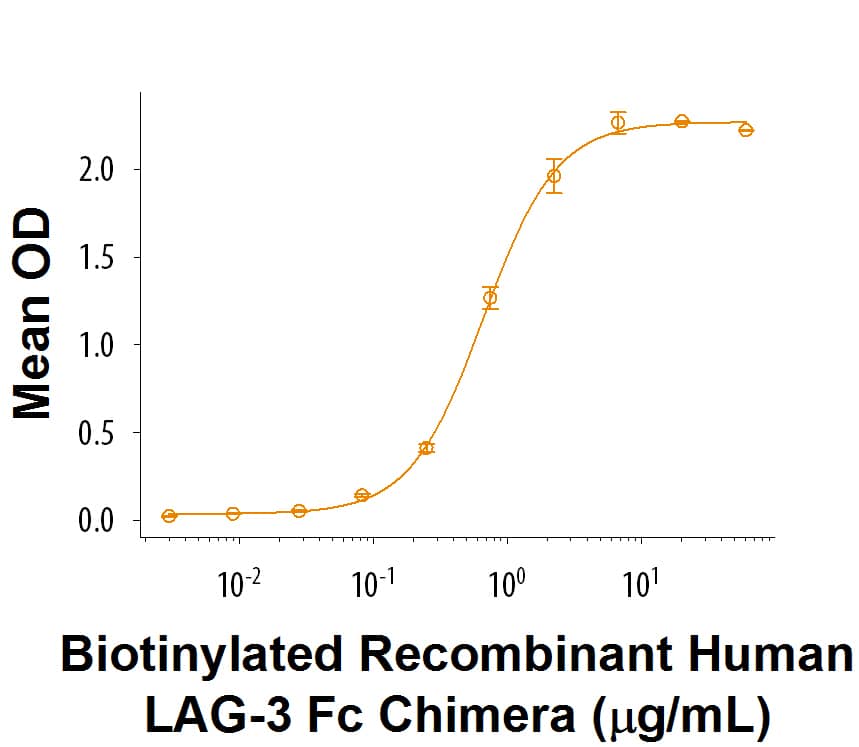
When Recombinant Human LSECtin/CLEC4G (Catalog # 2947-CL) is coated at 1 μg/mL, 100 uL/well, biotinylated recombinant LAG-3 Fc Chimera (Catalog# BT2319) binds with an ED50 of 0.2-2 μg/mL.
Scientific Data Images for Recombinant Human LAG-3 Fc Chimera Biotinylated Protein, CF
Recombinant Human LAG-3 Fc Chimera Biotinylated Protein Binding Activity
When Recombinant Human LSECtin/CLEC4G (Catalog # 2947-CL) is coated at 1 µg/mL, 100 µL/well, Biotinylated Recombinant Human LAG-3 Fc Chimera Biotinylated (Catalog # BT2319) binds with an ED50 of 0.2-2 µg/mL.Formulation, Preparation and Storage
BT2319
| Formulation | Lyophilized from a 0.2 μm filtered solution in PBS with Trehalose. |
| Reconstitution | Reconstitute at 200 μg/mL in PBS. |
| Shipping | The product is shipped at ambient temperature. Upon receipt, store it immediately at the temperature recommended below. |
| Stability & Storage | Use a manual defrost freezer and avoid repeated freeze-thaw cycles.
|
Background: LAG-3
LAG-3 (Lymphocyte activation gene-3), designated CD223, is a 70 kDa type I transmembrane protein that is a member of the immunoglobulin superfamily (IgSF) (1, 2). LAG-3 shares approximately 20% amino acid sequence homology with CD4, but has similar structure and binds to MHC class II with higher affinity, providing negative regulation of T cell receptor signaling (1, 2). Human LAG-3 cDNA encodes 525 amino acids (aa) that include a 28 aa signal sequence, a 422 aa extracellular domain (ECD) with four Ig-like domains, a transmembrane region and a highly charged cytoplasmic region. Within the ECD, human LAG-3 shares 70%, 67%, 76%, and 73% aa sequence identity with mouse, rat, porcine, and bovine LAG-3, respectively. LAG-3 is expressed on activated CD4+ and CD8+ T cells, NK cells, and plasmacytoid dendritic cells (pDC), but not on resting T cells (1-3). LAG-3 on activated CD4+CD25+ Treg cells plays a role in their suppressive activity (4). LAG-3 limits the expansion of activated T cells and pDC in response to selected stimuli (3-5). A soluble 54 kDa form, sLAG-3, can be shed by metalloproteinases ADAM10 and TACE/ADAM17 (6, 7). While monomeric sLAG-3 itself may be inactive, shedding allows for normal T cell activation by removing negative regulation (7). Binding of a homodimerized sLAG-3/Ig fusion protein to MHC class II molecules induces maturation of immature DC, and secretion of cytokines such as IFN-gamma and TNF-alpha by type 1 cytotoxic CD8+ T cells and NK cells (8, 9). sLAG-3/Ig has been used as a potential adjuvant to stimulate a cytotoxic anti-cancer immune response (9, 10). In mice, deletion of LAG-3 and another negative regulator, PD-1, facilitates anti-cancer response but also blocks self-tolerance and increases susceptibility to autoimmune diseases (11, 12). In humans, antibody-mediated down‑regulation of LAG-3 and PD-1 allows more effective control of chronic malaria, while in NOD (non‑obese diabetic) mice, deletion of LAG-3 alone accelerates diabetes (12-14).
References
- Triebel, F. et al. (1990) J. Exp. Med. 171:1393.
- Baixeras, E. et al. (1992) J. Exp. Med 176:327.
- Workman, C.J. et al. (2004) J. Immunol. 172:5450.
- Huang, C.T. et al. (2004) Immunity 21:503.
- Workman, C.J. et al. (2009) J. Immunol. 182:1885.
- Li, N. et al. (2004) J. Immunol. 173:6806.
- Li, N. et al. (2007) EMBO J. 26:494.
- Andreae, S. et al. (2003) Blood 102:2130.
- Brignone, C. et al. (2007) J. Immunol. 179:4202.
- Brignone, C. et al. (2010) J. Transl. Med. 8:71.
- Woo, S.R. et al. (2011) Cancer Res. 72:917.
- Okazaki, T. et al. (2011) J. Exp. Med. 208:395.
- Bettini, M. et al. (2011) J. Immunol. 187:3493.
- Butler, N.S. et al. (2012) Nat. Immunol. 13:188.
Long Name
Alternate Names
Gene Symbol
UniProt
Additional LAG-3 Products
Product Documents for Recombinant Human LAG-3 Fc Chimera Biotinylated Protein, CF
Product Specific Notices for Recombinant Human LAG-3 Fc Chimera Biotinylated Protein, CF
For research use only
