Recombinant Mouse CACHD1 Fc Chimera Protein, CF

R&D Systems, part of Bio-Techne | Catalog # 10754-CA

R&D Systems, part of Bio-Techne
Catalog #
Availability
Size / Price
Qty
Loading...
10754-CA-050

Key Product Details

Source

NS0

Accession #

Structure / Form

Disulfide-linked homodimer

Conjugate

Unconjugated

Applications

Bioactivity

Product Specifications

Source

Mouse myeloma cell line, NS0-derived mouse CACHD1 protein
Mouse CACHD1
(Asp52-Pro1109)
Accession # Q6PDJ1.1
IEGRMDP Mouse IgG2a
(Glu98-Lys330)
N-terminus C-terminus

Purity

>95%, by SDS-PAGE visualized with Silver Staining and quantitative densitometry by Coomassie® Blue Staining.

Endotoxin Level

<0.10 EU per 1 μg of the protein by the LAL method.

N-terminal Sequence Analysis

Asp52

Predicted Molecular Mass

145 kDa

SDS-PAGE

130-144 kDa, under reducing conditions.

Activity

Measured by its binding ability in a functional ELISA.
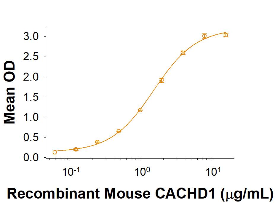
Recombinant Mouse CACHD1 Fc Chimera (Catalog # 10754-CA) binds Recombinant Human/Mouse Wnt-5a Biotinylated (Catalog # BT645) . The ED50 for this effect is 0.60-4.80 μg/mL.

Scientific Data Images for Recombinant Mouse CACHD1 Fc Chimera Protein, CF

Recombinant Mouse CACHD1 Fc Chimera Protein Binding Activity.

Recombinant Mouse CACHD1 Fc Chimera (Catalog # 10754-CA) binds Recombinant Human/Mouse Wnt-5a Biotinylated Protein (BT645). The ED50 for this effect is 0.60-4.80 μg/mL.

Recombinant Mouse CACHD1 Fc Chimera Protein SDS PAGE.

2 μg/lane of Recombinant Mouse CACHD1 Fc Chimera (Catalog # 10754-CA) was resolved with SDS-PAGE under reducing (R) and non-reducing (NR) conditions and visualized by Coomassie® Blue staining, showing bands at 130-144 kDa and 250-300 kDa, respectively.

Formulation, Preparation and Storage

10754-CA
Formulation Lyophilized from a 0.2 μm filtered solution in PBS.
Reconstitution Reconstitute at 500 μg/mL in PBS.
Shipping The product is shipped with polar packs. Upon receipt, store it immediately at the temperature recommended below.
Stability & Storage Use a manual defrost freezer and avoid repeated freeze-thaw cycles.
  • 12 months from date of receipt, -20 to -70 °C as supplied.
  • 1 month, 2 to 8 °C under sterile conditions after reconstitution.
  • 3 months, -70 °C under sterile conditions after reconstitution.

Background: CACHD1

CACHD1 (Ca2+ channel and chemotaxis receptor Domain Containing 1) is a alpha2 delta-Like Protein that modulates CaV3 voltage-gated calcium channel activity (1, 2). Mature Mouse CACHD1 consists of a 1060 amino acid (aa) extracellular domain (ECD), a 21 aa transmembrane segment, and a 158 aa cytoplasmic domain. The ECD contains a von Willebrand factor A (VWA) domain and two bacterial chemosensory-like cache domains. Within the ECD, mouse CACHD1 shares 97.5% and 99.7% aa sequence identity with human and rat CACHD1, respectively. Diseases associated with CACHD1 include Cercarial Dermatitis and Hypertropia. CACHD1 increases the presence of CaV3.1 at the cell surface and causes an increase in channel open probability (2). CACHD1 is a new activity-modifying protein for voltage-gated calcium channels (2). Such physiological actions may have implications in targeting diseases involving aberrant neuronal firing, as CaV3 channels have been proposed as therapeutic targets to combat pain and epilepsy (2). CACHD1 and MDGA1 are novel in vivo substrates for beta-site APP cleaving enzyme 1 (BACE1), suggesting that cleavage of both proteins may contribute to the numerous functions of BACE1 in the nervous system (3). BACE1 inhibition is a major therapeutic approach for Alzheimer disease (3). CACHD1 closely interacts with voltage-gated calcium channels of the N-type58 and T-type59 at the cell surface and enhances the densities of their calcium currents (3). BACE1 cleavage is a mechanism to control CACHD1's function in calcium signaling and synaptic transmission (3). Expression of CACHD1 increased both CaV2.2 currents and cell surface trafficking in both cell lines and neurons (4). CACHD1 competed with a2d-1 for binding to CaV2.2 and for its functional effects and can therefore inhibit responses to a2d-1 (4). CACHD1 is widely expressed in many tissues, including the brain, lungs, and small intestine (4). CACHD1 contains a VWA domain that has a disrupted metal ion-dependent adhesion site (MIDAS) motif, a sequence that is conserved in the human, rat, mouse, and zebrafish CACHD1 proteins (4). At the Wnt Signaling Gordon Conference in August 2019, Yvonne Jones (Oxford) presented an unpublished crystal structure of the transmembrane proteins CACHD1 in a complex between LRP6 and Fz5. Their data suggest that CACHD1 is an inhibitor of Wnt signaling.

References

  1. Cottrell, G.S. et al. (2018) J. Neurosci. 38:9186.
  2. Stephens, G.J. and G. S. Cottrell (2019) Channels (Austin) 13:120.
  3. Njavro, J.R. et al. (2020) The FASEB J. 34:2465.
  4. Dahimene, S. et al. (2018) Cell Reports 25:1610.

Long Name

Cache Domain Containing 1

Alternate Names

VWCD1

Entrez Gene IDs

57685 (Human); 320508 (Mouse)

Gene Symbol

CACHD1

UniProt

Additional CACHD1 Products

Product Documents for Recombinant Mouse CACHD1 Fc Chimera Protein, CF

Certificate of Analysis

To download a Certificate of Analysis, please enter a lot number in the search box below.

Note: Certificate of Analysis not available for kit components.

Product Specific Notices for Recombinant Mouse CACHD1 Fc Chimera Protein, CF

For research use only

Loading...
Loading...
Loading...