Goat anti-Canine IgG (H+L) Secondary Antibody [Alkaline Phosphatase]
Novus Biologicals, part of Bio-Techne | Catalog # NBP1-72567

Conjugate
Catalog #
Key Product Details
Species Reactivity
Canine
Applications
ELISA, Immunohistochemistry, Western Blot
Label
Alkaline Phosphatase
Antibody Source
Polyclonal Goat IgG
Concentration
Please see the vial label for concentration. If unlisted please contact technical services.
Product Specifications
Immunogen
Dog IgG whole molecule
Clonality
Polyclonal
Host
Goat
Isotype
IgG
Description
Store vial at 4C before opening. DO NOT FREEZE. This product is stable at 4C as an undiluted liquid. Dilute only prior to immediate use. Freezing alkaline phosphatase conjugates will result in a substantial loss of enzymatic activity.
This product was prepared from monospecific antiserum by immunoaffinity chromatography using Dog IgG coupled to agarose beads. Assay by immunoelectrophoresis resulted in a single precipitin arc against anti-Alkaline Phosphatase (calf intestine), anti-Goat Serum, Dog IgG and Dog Serum.
This product was prepared from monospecific antiserum by immunoaffinity chromatography using Dog IgG coupled to agarose beads. Assay by immunoelectrophoresis resulted in a single precipitin arc against anti-Alkaline Phosphatase (calf intestine), anti-Goat Serum, Dog IgG and Dog Serum.
Scientific Data Images for Goat anti-Canine IgG (H+L) Secondary Antibody [Alkaline Phosphatase]
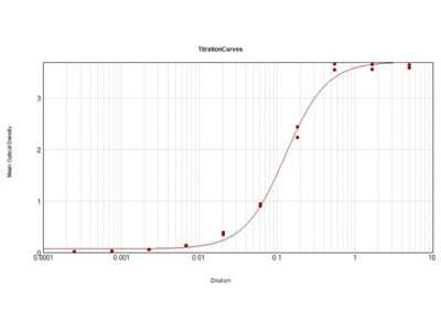
ELISA: Goat anti-Canine IgG (H+L) Secondary Antibody [Alkaline Phosphatase] [NBP1-72567] - ELISA Results of Goat anti-Canine IgG (H+L) Secondary antibody [Alkaline Phosphatase] tested against purified Dog IgG Alk Phos. Each well was coated in duplicate with 1.0 ug of Dog IgG. The working dilution is 1:8000. The starting dilution of antibody was 5ug/ml and the X-axis represents the Log10 of a 3-fold dilution. This titration is a 4-parameter curve fit where the IC50 is defined as the titer of the antibody. Assay performed using Alkaline Phosphatase Substrate ELISA Buffer and NPP Working Buffer.
Applications for Goat anti-Canine IgG (H+L) Secondary Antibody [Alkaline Phosphatase]
Application
Recommended Usage
ELISA
1:2000 - 1:10000
Immunohistochemistry
1:200 - 1:1000
Western Blot
1:500 - 1:2500
Application Notes
This product has been tested by ELISA. This product has been assayed against 1.0 ug of Dog IgG in a standard capture ELISA using pNPP p-nitrophenyl phosphate as a substrate for 30 minutes at room temperature. A working dilution of 1:1,000 to 1:5,000 of the reconstitution concentration is suggested for this product.
Formulation, Preparation, and Storage
Purification
Immunogen affinity purified
Formulation
0.05 M Tris Chloride, 0.15M Sodium Chloride, 0.001M Magnesium Chloride, 0.0001M Zinc Chloride, 50% (v/v) Glycerol; pH 8.0, 10 mg/mL Bovine Serum Albumin (BSA) - Immunoglobulin and Protease free
Preservative
0.01% Sodium Azide
Concentration
Please see the vial label for concentration. If unlisted please contact technical services.
Shipping
The product is shipped with polar packs. Upon receipt, store it immediately at the temperature recommended below.
Stability & Storage
Store at 4C. Do not freeze.
Background: IgG (H+L)
The 4 IgG subclasses, sharing 95% amino acid identity, include IgG1, IgG2, IgG3, and IgG4 for humans and IgG1, IgG2a, IgG2b, and IgG3 for mice. The relative abundance of each human subclass is 60% for IgG1, 32% for IgG2, 4% for IgG3, and 4% for IgG4. In an IgG deficiency, there may be a shortage of one or more subclasses (4).
References
1. Painter RH. (1998) Encyclopedia of Immunology (Second Edition). Elsevier. 1208-1211
2. Chapter 9 - Antibodies. (2012) Immunology for Pharmacy. Mosby 70-78
3. Schroeder H, Cavacini, L. (2010) Structure and Function of Immunoglobulins. J Allergy Clin Immunol. 125(2 0 2): S41-S52. PMID: 20176268
4. Vidarsson G, Dekkers G, Rispens T. (2014) IgG subclasses and allotypes: from structure to effector functions. Front Immunol. 5:520. PMID: 25368619
Additional IgG (H+L) Products
Product Documents for Goat anti-Canine IgG (H+L) Secondary Antibody [Alkaline Phosphatase]
Product Specific Notices for Goat anti-Canine IgG (H+L) Secondary Antibody [Alkaline Phosphatase]
This product is for research use only and is not approved for use in humans or in clinical diagnosis. Secondary Antibodies are guaranteed for 1 year from date of receipt.
Loading...
Loading...
Loading...
Loading...