What is Apoptosis?
Apoptosis is a coordinated and step-wise series of biochemical reactions resulting in the ordered disassembly of a cell within an organism. Defects in the apoptotic pathway that prevent cell death may lead to developmental abnormalities or unregulated tissue growth, as occurs in cancer. In contrast, pathological increases in apoptotic activity are hallmarks of several disease states including AIDS, neurodegenerative disorders, insulin-dependent diabetes, myocardial infarction, and atherosclerosis. Consequently, manipulation of the apoptotic process is essential to better understand the development of various diseases and to discover potential therapeutic targets.
Cytology of Apoptosis

Induction of apoptosis evokes several significant biomolecular and morphological changes, some of which are commonly used as markers of apoptosis. These events lead to major phenotypic alterations, highlighted in the figure above. Apoptosis concludes with the formation of apoptotic bodies, which are cleared by phagocytes or neighbouring cells.

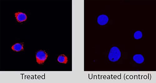
Immunocytochemical analysis of Caspase-3 expression in staurosporine treated vs untreated Jurkat cell line
Caspase-3 was detected in immersion fixed Jurkat human acute T cell leukemia cell line treated with Staurosporine (Catalog # 1285) using Rabbit Anti-Human Caspase-3 Monoclonal Antibody (Catalog # MAB7071) at 1 µg/mL for 3 hours at room temperature. Cells were stained using the NorthernLights™ 557-conjugated Anti-Rabbit IgG Secondary Antibody (red; Catalog # NL004) and counterstained with DAPI (blue). Specific staining was localized to cytoplasm.
Learn More About Apoptosis Signaling
Apoptosis occurs via a complex signaling cascade that triggers several significant changes and eventually leads to cell death. These molecular events can be used as potential biomarkers of apoptosis, including:
- Activation of apoptotic signaling cascades including the Bcl-2 family
- Phosphatidylserine Externalization
- Mitochondrial changes and the release of Cytochrome c
- Caspase cleavage and activation
- DNA fragmentation
Small molecule apoptosis activators and inhibitors are useful tools for manipulating and exploring apoptosis signaling.
Induce Apoptosis with Ease
Induce Apoptosis with Ease
Apoptosis inducing compounds exert pro-apoptotic effects via a variety of mechanisms including DNA cross-linking, inhibition of anti-apoptotic protein and caspase activation.
Simultaneously Detect Apoptosis-related Proteins
Simultaneously Detect Apoptosis-related Proteins
Rapid, sensitive, all-in-one kit for detecting relative expression of up to 35 apoptosis-related proteins in a single sample.
Simplify Apoptosis Detection
Simplify Apoptosis Detection
Choose from in situ detection of apoptosis in fixed frozen, paraffin-embedded, or plastic-embedded cells and tissues OR identify and quantify cells in suspension by flow cytometry.
Apoptosis Detection Methods
Apoptosis can be detected by a number of different methods, each of which has advantages and disadvantages. Best practice dictates that more than one method should be used to verify an apoptotic event. Find Protocols for Apoptosis Detection.

Timeline for Observing Apoptotic Events
Apoptosis is a dynamic process and determining the optimal time to measure the incidence of apoptosis or the activity of the molecules involved is critical for understanding the underlying mechanisms.
Several factors can influence the timing and should be considered in experimental design, including:
- cell type
- apoptotic stimulus
- the specific apoptotic mechanism
- experimental conditions.
Apoptotic Mechanisms May Vary with Cell Types
Several factors can affect how cells respond to an apoptosis-inducing agent and so it is important to understand the biological system under study.
- Primary cells may respond to death stimuli differently compared to immortalized cells.
- Cancer cell lines may respond differently from primary tumors or freshly explanted cells.
For instance, the human breast adenocarcinoma cell line, MCF-7, lacks Caspase 3, while primary neurons do not express Bcl-2 family member Bak, depending almost exclusively on Bax for mitochondria-mediated apoptosis.


