Exosomes and Other EVs in Neurodegenerative Diseases
Neurodegenerative diseases, like Alzheimer’s and Parkinson’s Disease, place a large burden on healthcare systems, with limited treatment options and no cure. Current methods of diagnosis rely on behavioral symptoms like memory loss and loss of motor functions. By the time an individual is symptomatic, there is irreparable damage to neurons and surrounding tissue. Given their accessibility in CSF and plasma, exosomes and other EVs are of great interest as a source of biomarkers for early detection of these diseases.
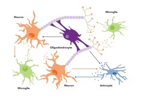
Exosomes and other EVs are involved in intercellular communication between all brain cells including neurons, astrocytes, microglia, and neural progenitor cells. EVs participate in many critical processes throughout the nervous system including neurogenesis, synaptic function, plasticity, and neuroinflammation.

EVs are involved in intercellular communication between all cell types in the nervous system. Arrows show flow of information from each cell type. Image adapted from Riva, P. et al. (2019) Emerging role of genetic alterations affecting exosome biology in neurodegenerative diseases. Int. J. Mol. Sci. 20 4113. PMID: 31450727. Licensed by CC License.
Exosomes and Other EVs in Alzheimer’s Disease
Alzheimer’s Disease (AD) is the most common form of dementia, marked by a progressive cognitive decline including memory loss and behavioral changes. According to the WHO, AD accounts for 60-70% of dementia cases. AD is characterized by amyloid-β (Aβ) accumulation in the extracellular space of neural tissues, as well as intracellular neurofibrillary tangles consisting of phosphorylated tau protein.

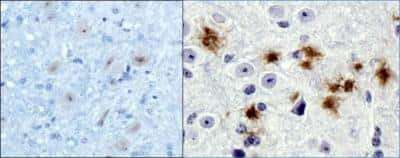
Immunohistochemistry (IHC) staining of β-amyloid in brain of wild-type (left) and 5xFAD (right) mouse using DAB with hematoxylin counterstain. Mouse Anti-Human beta Amyloid (MOAB-2) (Catalog # NBP2-13075) was used at 1:20 dilution in wild-type brain tissue and 1:400 dilution in 5xFAD mouse brain tissue. β-amyloid plaques indicated by blue arrows.
Extracellular Vesicles in AD Pathogenesis: Pathogenic or Protective?
The role of EVs in AD pathogenesis is not well defined, though there is consensus that exosomes are able to carry Aβ. However, there are conflicting reports about whether this association is protective or pathogenic. Some research shows a reduction in synaptic inflammation and increased disease pathology associated with an increase in EV-associated Aβ. Other research, however, shows exosomes protecting neurons transporting excess Aβ from neurons to microglia for degradation.
| Exosome Markers | Role in Alzheimer's Disease |
|---|---|
| Alix | Associated with Aβ plaques of AD brain, but not healthy controls |
| TSG101, VPS4A | Inhibition of transcription led to decreased spread of Aβ |
| Amyloid Precursor Protein (APP) | Precursor protein that is processed to Aβ |
| Aβ | Accumulation of plaques is associated with progressive cognitive decline |
| Tau, pTau (T181) | Major component of neurofibrillary tangles |
Early detection of AD could prove critical for intervention efforts to prevent or delay disease progression, so research is ongoing to identify biomarkers of AD to aid diagnosis. In addition to Aβ, proteins related to synaptic function are a high priority for biomarker research.
| Exosome Cargo | Expression in AD Patients | Present in Preclinical Disease; Associated with Disease State |
|---|---|---|
| Aβ42 | Increased in plasma | EV-associated levels can predict risk of disease progression |
| TAR DNA-binding protein 43 (TDP43) | Increased in plasma | No correlation with cognitive function or other neurological symptoms |
| Synaptophysin | Decreased in plasma | Already decreased in preclinical state; correlated with cognitive decline |
| Synaptotagmin | Decreased in plasma | Already decreased in preclinical state; correlated with cognitive decline |
| 25-kDa synaptosomal-associated protein (SNAP-25) | Decreased in plasma | Can be detected several years before clinical onset of AD |
| GAP43 | Decreased in plasma | Can be detected several years before clinical onset of AD |
| Neurogranin | Increased in CSF, decreased in plasma | Present in preclinical state. Potentially predictive of disease progression |
Exosomes and Other EVs in Parkinson’s Disease
Parkinson’s Disease (PD) is another common neurodegenerative disease, characterized by the loss of dopaminergic neurons, primarily in the substantia nigra.
PD is defined by the presence of α-synuclein aggregates (Lewy bodies) and accumulation of defective mitochondria, leading to neuronal death. Exosomes, specifically L1CAM-expressing neuronal-derived exosomes (NDEs) have been implicated in the pathological transport of α-synuclein in the brain. Diagnosis of PD relies on neurological examinations and presentation of symptoms. The most consistent exosome biomarker in PD patients is increased levels of NDE-associated α-synuclein in plasma. Other targets associated with plasma NDEs in PD include Park7/DJ1 and clusterin.
Currently, there is no cure for PD, although motor symptoms can be temporarily alleviated with Levodopa which is converted to Dopamine in neurons. As new therapies concentrate on neuroprotective and disease-modifying strategies, early detection through the identification of exosome markers for Parkinson’s disease is a critical research focus.
Beatriz M, et al. (2021) Exosomes: innocent bystanders or critical culprits in neurodegenerative diseases. Front Cell Dev Biol 9 635104. https://doi.org/10.3389/fcell.2021.635104.
Chung IM, et al. (2020) Exosomes: Current use and future applications. Clin Chim Acta 500 226. https://doi.org/10.1016/j.cca.2019.10.022.
D’Anca M, et al. (2019) Exosome determinants of physiological aging and age-related neurodegenerative diseases. Front Aging Neurosci 11 232. https://doi.org/10.3389/FNAGI.2019.00232.
Frühbeis C, et al. (2012) Emerging roles of exosomes in neuron-glia communication. Front Physiol 3 119. https://doi.org/10.3389/fphys.2012.00119.
Liu W, et al. (2020) Neurogranin as a cognitive biomarker in cerebrospinal fluid and blood exosomes for Alzheimer’s disease and mild cognitive impairment. Transl Psychiatry 10 125. https://doi.org/10.1038/s41398-020-0801-2.
Pulliam L, et al. (2019) Plasma neuronal exosomes serve as biomarkers of cognitive impairment in HIV infection and Alzheimer’s disease. J Neurovirol 25 702. https://doi.org/10.1007/s13365-018-0695-4.
Riva P, et al. (2019) Emerging role of genetic alterations affecting exosome biology in neurodegenerative diseases. Int J Mol Sci 20 4113. https://doi.org/10.3390/IJMS20174113.