Caspase-1 Antibody (14F468) - BSA Free
Novus Biologicals, part of Bio-Techne | Catalog # NB100-56565


Conjugate
Catalog #
Forumulation
Catalog #
Key Product Details
Validated by
Biological Validation
Species Reactivity
Validated:
Human, Mouse, Rat
Cited:
Human, Mouse, Rat
Applications
Validated:
Immunocytochemistry/ Immunofluorescence, Immunohistochemistry, Immunohistochemistry-Frozen, Immunohistochemistry-Paraffin, Simple Western, Western Blot
Cited:
Block/Neutralize, ELISA, IF/IHC, Immunocytochemistry/ Immunofluorescence, Immunohistochemistry-Frozen, Immunohistochemistry-Paraffin, Western Blot
Label
Unconjugated
Antibody Source
Monoclonal Mouse IgG1 kappa Clone # 14F468
Format
BSA Free
Concentration
1.0 mg/ml
Product Specifications
Immunogen
Caspase-1 Antibody (14F468) was developed against two synthetic peptides from the human Caspase-1 protein (amino acids 371-390 and 31-45) [UniProt P29466].
Reactivity Notes
Immunogen's sequence similarity with other species: Porcine/Pig (85%), Equine/Horse (80%), Canine (70%). Rat reactivity reported in scientific literature (PMID: 22133203).
Localization
Cytoplasm, Nucleus
Specificity
Caspase-1 Antibody (14F468) will recognize full-length Caspase-1 and cleaved Caspase-1 forms that retain amino acids 371-390 of the Caspase-1 protein.
Clonality
Monoclonal
Host
Mouse
Isotype
IgG1 kappa
Theoretical MW
45.2 kDa.
Disclaimer note: The observed molecular weight of the protein may vary from the listed predicted molecular weight due to post translational modifications, post translation cleavages, relative charges, and other experimental factors.
Disclaimer note: The observed molecular weight of the protein may vary from the listed predicted molecular weight due to post translational modifications, post translation cleavages, relative charges, and other experimental factors.
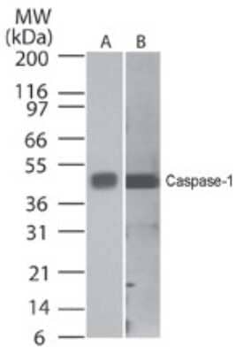
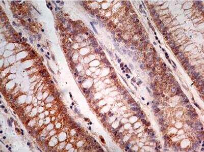
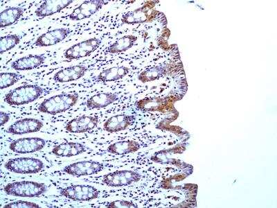
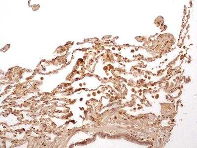
Scientific Data Images for Caspase-1 Antibody (14F468) - BSA Free
Detection of Caspase-1 in HeLa Cell Lysate by Simple Western
Simple Western lane view shows a specific band for Caspase 1 in 1.0 mg/ml of HeLa lysate. This experiment was performed under reducing conditions using the 12-230 kDa separation system.Immunohistological Staining of Caspase-1 in Control and OHT-Injured Mouse Eyes
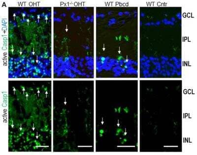
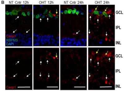
Activity of Casp1 in OHT-injured and normotensive control eyes. (A) Casp1 was detected by intraocular injection FLICA660-labeled substrate (green) in vivo 24 h after injury. Bright labeling (arrows) is evident in cells in the GCL and inner nuclear layer (INL) layers of the OHT-challenged retinas, a diffuse labeling of cell processes located in the IPL. Casp1 activity is diminished in Panx1-/- (Px1-/- OHT) retinas and WT retinas treated with probenecid (WT/Pbcd) at 12 h postinjury. Image collected and cropped by CiteAb from the following publication (https://www.frontiersin.org/article/10.3389/fnmol.2019.00036/full), licensed under a CC-BY license.Western Blot Analysis of Caspase-1 in Infected Mouse BMMs
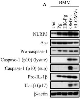
P. gingivalis and its OMVs differentially induce inflammasome signaling and pyroptosis in murine macrophages. BMM were infected as before (2 h at MOI of 25:1, see Materials and Methods) with viable P. gingivalis (Pg), heat-killed-Pg (HK-Pg), OMVs, or heat-inactivated-OMVs (HI-OMVs) and the activation of inflammasome components in the lysates [or supernatants (sup) where indicated] measured after 24 h by Western blot; beta-actin serves as a loading control throughout. Western blot data are representative of at least three independent experiments. Image collected and cropped by CiteAb from the following publication (https://journal.frontiersin.org/article/10.3389/fcimb.2017.00351/full), licensed under a CC-BY license.Applications for Caspase-1 Antibody (14F468) - BSA Free
Application
Recommended Usage
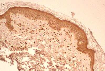
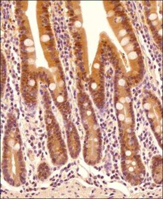
Immunohistochemistry
1:100 - 1:500
Immunohistochemistry-Frozen
reported in scientific literature (PMID 30930743)
Immunohistochemistry-Paraffin
1:10-1:500
Simple Western
1:50
Western Blot
0.5-2 ug/ml
Application Notes
Staining of formalin-fixed tissues is enhanced by boiling tissue sections in 10 mM sodium citrate buffer, pH 6.0 for 10-20 min followed by cooling at RT for 20 min.
In Simple Western only 10 - 15 uL of the recommended dilution is used per data point.
See Simple Western Antibody Database for Simple Western validation: Tested in HeLa lysate 1.0 mg/mL, separated by Size, antibody dilution of 1:50, apparent MW was 51 kDa. Separated by Size-Wes, Sally Sue/Peggy Sue.
Formulation, Preparation, and Storage
Purification
Protein G purified
Formulation
PBS
Format
BSA Free
Preservative
0.02% Sodium Azide
Concentration
1.0 mg/ml
Shipping
The product is shipped with polar packs. Upon receipt, store it immediately at the temperature recommended below.
Stability & Storage
Store at 4C short term. Aliquot and store at -20C long term. Avoid freeze-thaw cycles.
Background: Caspase-1
Given the role of IL-1beta in inflammation, it makes sense that many diseases and pathologies have been associated with dysregulation of caspase-1 activation and the inflammasome (3, 4). The inflammasome is a multiprotein complex comprised of Nod-like receptor (NLR) family members and the adapter ASC (apoptosis-associated speck-like protein containing a CARD) which are crucial for capase-1 activation (3-5). For instance, the neuronal apoptosis inhibitory protein (NAIP)/NLRC4 inflammasome has been associated with colorectal cancer, breast cancer, and glioma pathogenesis (5). Caspase-1 activation and mutations in the inflammasome have also been linked to Chron's disease and Alzheimer's disease (4). In addition to immune and inflammatory related disorder, the inflammasome has been linked to metabolic and obesity related disorders including diabetes and cardiovascular disease (6). Finally, caspase-1 deficient mice exhibit enhanced epithelial cell proliferation in the colon, increased tumor formation, and reduced apoptosis (1). A more thorough understanding of the inflammasome-caspase-1 signaling pathway will be important for understanding disease pathology and potential therapeutic development.
Alternative names for caspase-1 includes CASP1, CASP1 nirs variant 1, EC 3.4.22.36, ICE, IL-1 beta-converting enzyme, IL1BC, IL1BCE, IL1B-converstase, interleukin-1 beta convertase, and p45.
References
1. Shalini, S., Dorstyn, L., Dawar, S., & Kumar, S. (2015). Old, new and emerging functions of caspases. Cell death and differentiation. https://doi.org/10.1038/cdd.2014.216
2. Chang, H. Y., & Yang, X. (2000). Proteases for cell suicide: functions and regulation of caspases. Microbiology and molecular biology reviews: MMBR. https://doi.org/10.1128/mmbr.64.4.821-846.2000
3. Vanaja, S. K., Rathinam, V. A., & Fitzgerald, K. A. (2015). Mechanisms of inflammasome activation: recent advances and novel insights. Trends in cell biology. https://doi.org/10.1016/j.tcb.2014.12.009
4. Franchi, L., Eigenbrod, T., Munoz-Planillo, R., & Nunez, G. (2009). The inflammasome: a caspase-1-activation platform that regulates immune responses and disease pathogenesis. Nature immunology. https://doi.org/10.1038/ni.1703
5. Kay, C., Wang, R., Kirkby, M., & Man, S. M. (2020). Molecular mechanisms activating the NAIP-NLRC4 inflammasome: Implications in infectious disease, autoinflammation, and cancer. Immunological reviews. https://doi.org/10.1111/imr.12906
6. Pham, D., Park, P. (2020). Recent insights on modulation of inflammasomes by adipokines: a critical event for the pathogenesis of obesity and metabolism-associated diseases. Archives of Pharmacal Research. https://doi.org/10.1007/s12272-020-01274-7
Alternate Names
CASP1, Caspase1, ICE
Gene Symbol
CASP1
UniProt
Additional Caspase-1 Products
Product Documents for Caspase-1 Antibody (14F468) - BSA Free
Product Specific Notices for Caspase-1 Antibody (14F468) - BSA Free
This product is for research use only and is not approved for use in humans or in clinical diagnosis. Primary Antibodies are guaranteed for 1 year from date of receipt.
Loading...
Loading...
Loading...
Loading...
Loading...


![Western Blot: Caspase-1 Antibody (14F468) - BSA Free [NB100-56565] - Caspase-1 Antibody (14F468) - BSA Free](https://resources.bio-techne.com/images/products/nb100-56565_mouse-monoclonal-caspase-1-antibody-14f468-310202415175246.jpg)
![Western Blot: Caspase-1 Antibody (14F468) - BSA Free [NB100-56565] - Caspase-1 Antibody (14F468) - BSA Free](https://resources.bio-techne.com/images/products/nb100-56565_mouse-monoclonal-caspase-1-antibody-14f468-310202415345329.jpg)
![Western Blot: Caspase-1 Antibody (14F468) - BSA Free [NB100-56565] - Caspase-1 Antibody (14F468) - BSA Free](https://resources.bio-techne.com/images/products/nb100-56565_mouse-monoclonal-caspase-1-antibody-14f468-310202415384197.jpg)
![Western Blot: Caspase-1 Antibody (14F468) - BSA Free [NB100-56565] - Caspase-1 Antibody (14F468) - BSA Free](https://resources.bio-techne.com/images/products/nb100-56565_mouse-monoclonal-caspase-1-antibody-14f468-31020241539719.jpg)
![Western Blot: Caspase-1 Antibody (14F468) - BSA Free [NB100-56565] - Caspase-1 Antibody (14F468) - BSA Free](https://resources.bio-techne.com/images/products/nb100-56565_mouse-monoclonal-caspase-1-antibody-14f468-310202415392595.jpg)
![Western Blot: Caspase-1 Antibody (14F468) - BSA Free [NB100-56565] - Caspase-1 Antibody (14F468) - BSA Free](https://resources.bio-techne.com/images/products/nb100-56565_mouse-monoclonal-caspase-1-antibody-14f468-310202415175211.jpg)
![Western Blot: Caspase-1 Antibody (14F468) - BSA Free [NB100-56565] - Caspase-1 Antibody (14F468) - BSA Free](https://resources.bio-techne.com/images/products/nb100-56565_mouse-monoclonal-caspase-1-antibody-14f468-310202415395949.jpg)
![Western Blot: Caspase-1 Antibody (14F468) - BSA Free [NB100-56565] - Caspase-1 Antibody (14F468) - BSA Free](https://resources.bio-techne.com/images/products/nb100-56565_mouse-monoclonal-caspase-1-antibody-14f468-31020241534326.jpg)
![Western Blot: Caspase-1 Antibody (14F468) - BSA Free [NB100-56565] - Caspase-1 Antibody (14F468) - BSA Free](https://resources.bio-techne.com/images/products/nb100-56565_mouse-monoclonal-caspase-1-antibody-14f468-31020241612614.jpg)
![Western Blot: Caspase-1 Antibody (14F468) - BSA Free [NB100-56565] - Caspase-1 Antibody (14F468) - BSA Free](https://resources.bio-techne.com/images/products/nb100-56565_mouse-monoclonal-caspase-1-antibody-14f468-310202415541952.jpg)
![Western Blot: Caspase-1 Antibody (14F468) - BSA Free [NB100-56565] - Caspase-1 Antibody (14F468) - BSA Free](https://resources.bio-techne.com/images/products/nb100-56565_mouse-monoclonal-caspase-1-antibody-14f468-310202415541990.jpg)