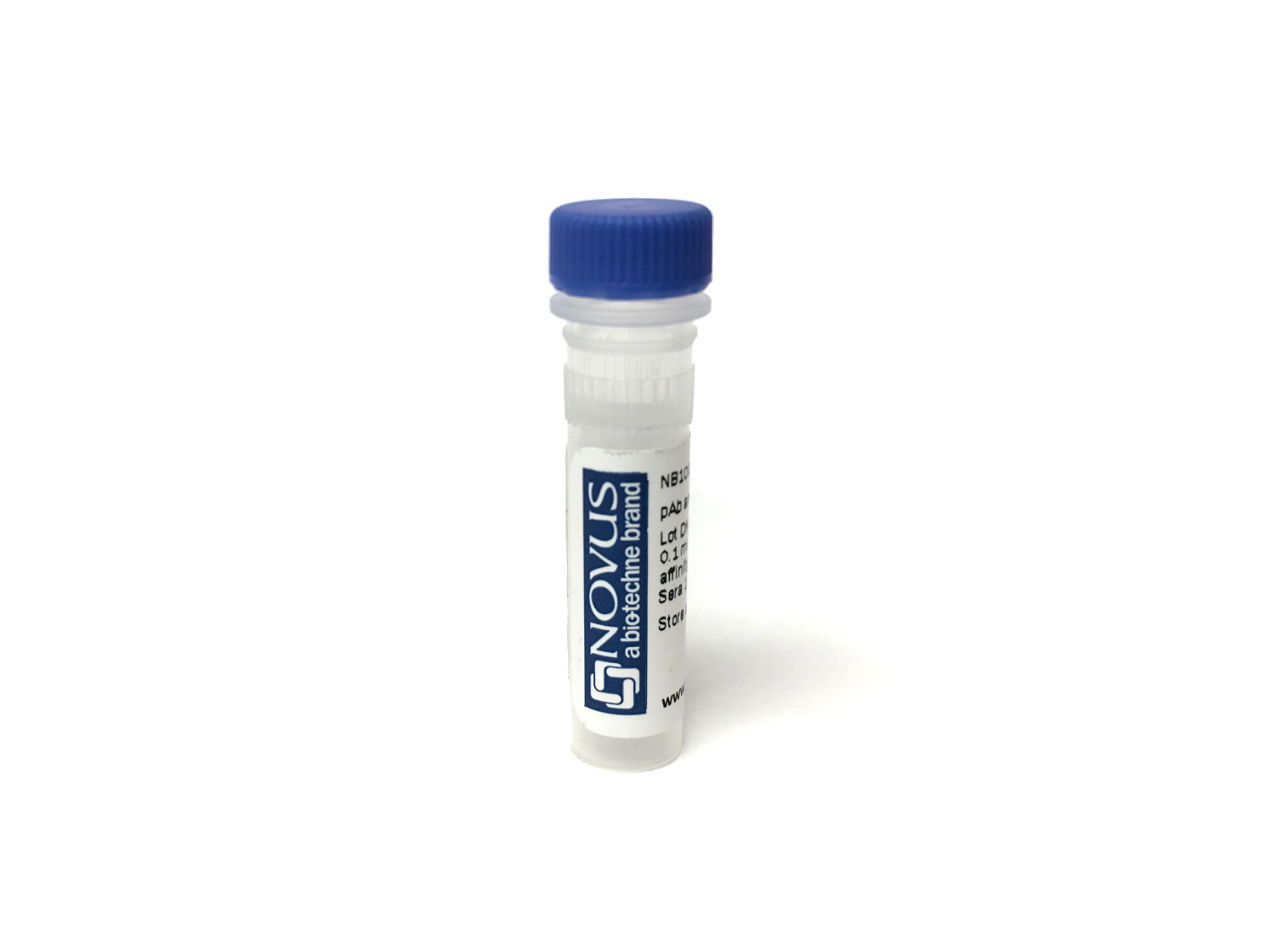
CD31/PECAM-1 Antibody (JC/70A) [DyLight 755]
Novus Biologicals, part of Bio-Techne | Catalog # NB600-562IR
Clone JC/70A was used by HLDA to establish CD designation.


Conjugate
Catalog #
Key Product Details
Species Reactivity
Human, Mouse, Feline, Rabbit
Applications
Dual RNAscope ISH-IHC, ELISA, Flow (Cell Surface), Flow Cytometry, Immunocytochemistry/ Immunofluorescence, Immunohistochemistry, Immunohistochemistry-Frozen, Immunohistochemistry-Paraffin, Knockout Validated, Western Blot
Label
DyLight 755 (Excitation = 754 nm, Emission = 776 nm)
Antibody Source
Monoclonal Mouse IgG1 kappa Clone # JC/70A
Concentration
Please see the vial label for concentration. If unlisted please contact technical services.
Product Specifications
Immunogen
This CD31/PECAM-1 Antibody (JC/70A) was developed against a membrane preparation of a spleen from a patient with hairy cell leukemia.
Reactivity Notes
Rabbit (PMID: 21533193) and Mouse (PMID: 29700126) reactivity reported in scientific literature.
Localization
Cell membrane.
Clonality
Monoclonal
Host
Mouse
Isotype
IgG1 kappa
Theoretical MW
82.5 kDa.
Disclaimer note: The observed molecular weight of the protein may vary from the listed predicted molecular weight due to post translational modifications, post translation cleavages, relative charges, and other experimental factors.
Disclaimer note: The observed molecular weight of the protein may vary from the listed predicted molecular weight due to post translational modifications, post translation cleavages, relative charges, and other experimental factors.
Applications for CD31/PECAM-1 Antibody (JC/70A) [DyLight 755]
Application
Recommended Usage
Dual RNAscope ISH-IHC
Optimal dilutions of this antibody should be experimentally determined.
ELISA
Optimal dilutions of this antibody should be experimentally determined.
Flow (Cell Surface)
Optimal dilutions of this antibody should be experimentally determined.
Flow Cytometry
Optimal dilutions of this antibody should be experimentally determined.
Immunocytochemistry/ Immunofluorescence
Optimal dilutions of this antibody should be experimentally determined.
Immunohistochemistry
Optimal dilutions of this antibody should be experimentally determined.
Immunohistochemistry-Frozen
Optimal dilutions of this antibody should be experimentally determined.
Immunohistochemistry-Paraffin
Optimal dilutions of this antibody should be experimentally determined.
Knockout Validated
Optimal dilutions of this antibody should be experimentally determined.
Western Blot
Optimal dilutions of this antibody should be experimentally determined.
Application Notes
Optimal dilution of this antibody should be experimentally determined.
Formulation, Preparation, and Storage
Purification
Protein G purified
Formulation
50mM Sodium Borate
Preservative
0.05% Sodium Azide
Concentration
Please see the vial label for concentration. If unlisted please contact technical services.
Shipping
The product is shipped with polar packs. Upon receipt, store it immediately at the temperature recommended below.
Stability & Storage
Store at 4C in the dark.
Background: CD31/PECAM-1
PECAM's intracellular cytoplasmic domain consists of a sequence of 118 amino acids and contains serine and tyrosine (also referred to as immunoreceptor tyrosine-based inhibitory motifs-ITIMs) residues, which may be phosphorylated upon cellular stimulation (3). ITIMs are phosphorylated by Src-family kinases and non-Src family kinases (e.g., Csk), leading to a conformational change which supports interactions with Src homology 2 (SH2) domain containing proteins such as protein-tyrosine phosphatase, SHP-2 (1,2). Formation of SHP-2/PECAM-1 complexes induces endothelial cell migration through the dephosphorylation of focal adhesion kinase and regulation of RhoA activity (1). Signaling downstream of ITIM tyrosine phosphorylations also plays a role in PECAM's anti-apoptotic activity, a function which is independent of its interaction with SHP-2. In platelets and leukocytes, phosphorylation of PECAM's cytosolic domain is inhibitory, preventing their activation.
References
1. Lertkiatmongkol, P., Liao, D., Mei, H., Hu, Y., & Newman, P. J. (2016). Endothelial functions of PECAM-1 (CD31). Current Opinion in Hematology. https://doi.org/10.1097/MOH.0000000000000239.Endothelial
2. Privratsky, J. R., & Newman, P. J. (2014). PECAM-1: Regulator of endothelial junctional integrity. Cell and Tissue Research. https://doi.org/10.1007/s00441-013-1779-3
3. Newman, P. J., & Newman, D. K. (2003). Signal transduction pathways mediated by PECAM-1: New roles for an old molecule in platelet and vascular cell biology. Arteriosclerosis, Thrombosis, and Vascular Biology. https://doi.org/10.1161/01.ATV.0000071347.69358.D9
Long Name
Platelet Endothelial Cell Adhesion Molecule 1
Alternate Names
CD31, EndoCAM, PECA1, PECAM-1, PECAM1
Gene Symbol
PECAM1
Additional CD31/PECAM-1 Products
Product Documents for CD31/PECAM-1 Antibody (JC/70A) [DyLight 755]
Product Specific Notices for CD31/PECAM-1 Antibody (JC/70A) [DyLight 755]
DyLight (R) is a trademark of Thermo Fisher Scientific Inc. and its subsidiaries.
This product is for research use only and is not approved for use in humans or in clinical diagnosis. Primary Antibodies are guaranteed for 1 year from date of receipt.
Loading...
Loading...
Loading...
Loading...
Loading...
Loading...