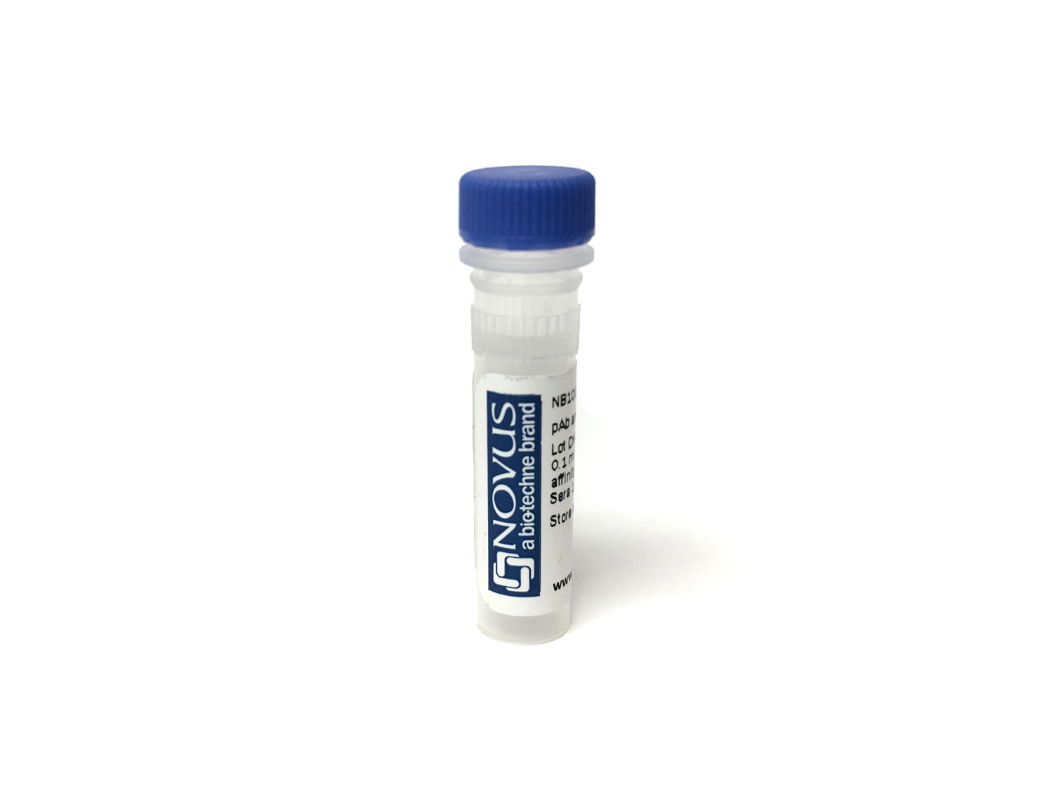
CD8 Antibody (53-6.7) [Biotin]
Novus Biologicals, part of Bio-Techne | Catalog # NBP1-49045B


Conjugate
Catalog #
Key Product Details
Species Reactivity
Mouse, Rat
Applications
Cell depletion, CyTOF-ready, Flow Cytometry, Immunocytochemistry/ Immunofluorescence, Immunohistochemistry, Immunohistochemistry-Frozen, Immunohistochemistry-Paraffin, Immunoprecipitation, Inhibition of T Cell Function
Label
Biotin
Antibody Source
Monoclonal Rat IgG2a Kappa Clone # 53-6.7
Concentration
Please see the vial label for concentration. If unlisted please contact technical services.
Product Specifications
Immunogen
CD8 Antibody (53-6.7) was developed against mouse thymus or spleen.
Localization
Most thymocytes, T cell subset, some NK cells
Clonality
Monoclonal
Host
Rat
Isotype
IgG2a Kappa
Applications for CD8 Antibody (53-6.7) [Biotin]
Application
Recommended Usage
Cell depletion
Optimal dilutions of this antibody should be experimentally determined.
CyTOF-ready
Optimal dilutions of this antibody should be experimentally determined.
Flow Cytometry
Optimal dilutions of this antibody should be experimentally determined.
Immunocytochemistry/ Immunofluorescence
Optimal dilutions of this antibody should be experimentally determined.
Immunohistochemistry
Optimal dilutions of this antibody should be experimentally determined.
Immunohistochemistry-Frozen
Optimal dilutions of this antibody should be experimentally determined.
Immunohistochemistry-Paraffin
Optimal dilutions of this antibody should be experimentally determined.
Immunoprecipitation
Optimal dilutions of this antibody should be experimentally determined.
Inhibition of T Cell Function
Optimal dilutions of this antibody should be experimentally determined.
Application Notes
Optimal dilution of this antibody should be experimentally determined.
Formulation, Preparation, and Storage
Purification
Protein A or G purified
Formulation
PBS
Preservative
0.05% Sodium Azide
Concentration
Please see the vial label for concentration. If unlisted please contact technical services.
Shipping
The product is shipped with polar packs. Upon receipt, store it immediately at the temperature recommended below.
Stability & Storage
Store at 4C in the dark.
Background: CD8
Given its role in the immune system, CD8-deficiency in T-cells is a hallmark of many diseases and pathologies (8-10). Specifically, CD8+ T-cell deficiency is prevalent in chronic autoimmune diseases including multiple sclerosis, rheumatoid arthritis, ulcerative colitis, Crohn's disease, type 1 diabetes mellitus, and Graves' disease (8). Furthermore, cancers or chronic infection can lead to CD8 T-cell exhaustion as the continual antigen presentation and inflammatory signals eventually cause the CD8+ T-cells to lose functionality (9, 10). However, animal models and clinical studies have suggested that T-cells are capable of being reinvigorated using inhibitory receptor blockade resulting in better disease outcomes and these exhausted T-cells may be a potential therapeutic target (9, 10).
Alternative names for CD8 includes CD antigen: CD8a, CD8 antigen, alpha polypeptide (p32), CD8a molecule, CD8A, Leu2 T-lymphocyte antigen, LEU2, MAL, OKT8 T-cell antigen, p32, T cell co-receptor, T8 T-cell antigen, T-cell antigen Leu2, T-cell surface glycoprotein CD8 alpha chain, and T-lymphocyte differentiation antigen T8/Leu-2.
References
1. Littman D. R. (1987). The structure of the CD4 and CD8 genes. Annual review of immunology. https://doi.org/10.1146/annurev.iy.05.040187.003021
2. Naeim F. (2008). Chapter 2- Principles of Immunophenotyping. Hematopathology. https://doi.org/10.1016/B978-0-12-370607-2.00002-8.
3. Gao, G. F., & Jakobsen, B. K. (2000). Molecular interactions of coreceptor CD8 and MHC class I: the molecular basis for functional coordination with the T-cell receptor. Immunology today. https://doi.org/10.1016/s0167-5699(00)01750-3
4. UniProt (P01732)
5. UniProt (P01731)
6. Kappes D. J. (2007). CD4 and CD8: hogging all the Lck. Immunity. https://doi.org/10.1016/j.immuni.2007.11.002
7. Gangadharan, D., & Cheroutre, H. (2004). The CD8 isoform CD8alphaalpha is not a functional homologue of the TCR co-receptor CD8alphabeta. Current opinion in immunology. https://doi.org/10.1016/j.coi.2004.03.015
8. Pender M. P. (2012). CD8+ T-Cell Deficiency, Epstein-Barr Virus Infection, Vitamin D Deficiency, and Steps to Autoimmunity: A Unifying Hypothesis. Autoimmune diseases. https://doi.org/10.1155/2012/189096
9. Kurachi M. (2019). CD8+ T cell exhaustion. Seminars in immunopathology. https://doi.org/10.1007/s00281-019-00744-5
10. Hashimoto, M., Kamphorst, A. O., Im, S. J., Kissick, H. T., Pillai, R. N., Ramalingam, S. S., Araki, K., & Ahmed, R. (2018). CD8 T Cell Exhaustion in Chronic Infection and Cancer: Opportunities for Interventions. Annual review of medicine. https://doi.org/10.1146/annurev-med-012017-043208
Alternate Names
CD8, CD8A
Gene Symbol
CD8A
Additional CD8 Products
Product Documents for CD8 Antibody (53-6.7) [Biotin]
Product Specific Notices for CD8 Antibody (53-6.7) [Biotin]
This product is for research use only and is not approved for use in humans or in clinical diagnosis. Primary Antibodies are guaranteed for 1 year from date of receipt.
Loading...
Loading...
Loading...
Loading...
Loading...