GFP Antibody [DyLight 488]
Novus Biologicals, part of Bio-Techne | Catalog # NBP1-69969


Forumulation
Catalog #
Key Product Details
Species Reactivity
Non-species specific
Applications
Validated:
Dot Blot, ELISA, Fluorophore-linked immunosorbent assay, Immunocytochemistry/ Immunofluorescence, Knockdown Validated, Knockout Validated, Western Blot
Cited:
Immunocytochemistry/ Immunofluorescence
Label
DyLight 488 (Excitation = 493 nm, Emission = 518 nm)
Antibody Source
Polyclonal Goat IgG
Concentration
LYOPH mg/ml
Product Specifications
Immunogen
The immunogen is a Green Fluorescent Protein (GFP) fusion protein corresponding to the full length amino acid sequence (246aa) derived from the jellyfish Aequorea victoria. (Uniprot: P42212)
Reactivity Notes
No reaction was observed against Human, or Rat serum proteins. Known Cross Reactivity: rGFP. YFP differs from GFP due to a mutation at Thr203Tyr; antibodies raised against full-length GFP should also detect YFP and other variants. Reactivity in transgenic mice with GFP. Reactivity in human cell lines transfected will a GFP construct.
Specificity
No reaction was observed against Human, Mouse or Rat serum proteins.
Clonality
Polyclonal
Host
Goat
Isotype
IgG
Description
Store vial at 4C prior to restoration. For extended storage aliquot contents and freeze at -20C or below. Avoid cycles of freezing and thawing. Centrifuge product if not completely clear after standing at room temperature. This product is stable for several weeks at 4C as an undiluted liquid. Dilute only prior to immediate use.
GFP Dylight(TM) 488 Conjugated Antibody was prepared from monospecific antiserum by immunoaffinity chromatography using Green Fluorescent Protein (Aequorea victoria) coupled to agarose beads followed by solid phase adsorption(s) to remove any unwanted reactivities. Assay by immunoelectrophoresis resulted in a single precipitin arc against anti-Goat Serum and purified and partially purified Green Fluorescent Protein (Aequorea victoria)
GFP Dylight(TM) 488 Conjugated Antibody was prepared from monospecific antiserum by immunoaffinity chromatography using Green Fluorescent Protein (Aequorea victoria) coupled to agarose beads followed by solid phase adsorption(s) to remove any unwanted reactivities. Assay by immunoelectrophoresis resulted in a single precipitin arc against anti-Goat Serum and purified and partially purified Green Fluorescent Protein (Aequorea victoria)
Scientific Data Images for GFP Antibody [DyLight 488]
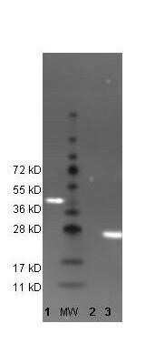
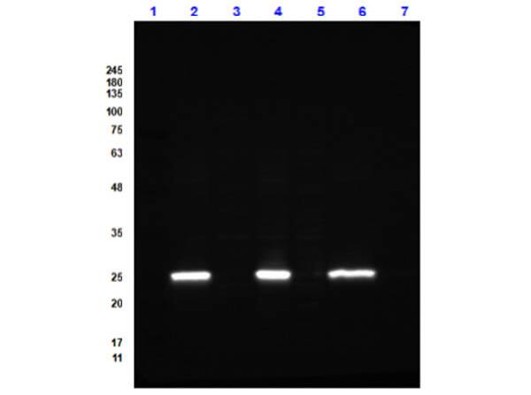
Western Blot Detection of GFP Using DyLight 488 Conjugated Antibody
Lane 1: His-Sumo-GFP. Lane: Molecular Weight. Lane 2: Beta-Galactosidase (negative control). Lane 3: recombinant GFP control protein. Load: 35 ug per lane. Primary antibody: none. Secondary antibody: DyLight (TM) 488 conjugated anti-GFP goat secondary antibody at 1:5,000. Block for2 hr at RT. Predicted/Observed size: 27kDa/54kDa, 27kDa for rGFP/~45kDa His-Sumo-GFP.Comparison of Fluorescence Absorption and Emission Spectra for DyLight 488 and Alexa Fluor 488 Conjugates
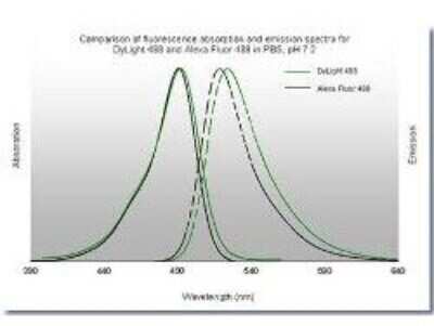
Comparison of fluorescence absorption and emission spectra for DyLight (TM) 488 and Alexa Fluor 488 in PBS, pH7.2. The emission spectra for this DyLight (TM) conjugate match the principle output wavelengths of most common fluorescence instrumentation.Immunofluorescent Staining of Human Breast Carcinoma Tissue Using DyLight 488 Conjugated GFP Antibody
Tissue: human breast carcinoma. Fixation: 0.5% PFA. Antigen retrieval: not required. Primary antibody: Anti-Histone and Anti-Tubulin antibody at 10 ug/mL for 1 h at RT. Secondary antibody: DyLight (R) 488 conjugate and DyLight (R) 549 conjugate goat secondary antibody at 1:10,000 for 45 min at RT. Localization: Histone is nuclear and Tubulin is cytoplasmic. Staining: Anti-Histone detection using a DyLight (R) 488 conjugate (green) fluorescent signal and Anti-Tubulin was detected using a DyLight (R) 549 conjugate (red) fluorescent signal. Nuclei were counter-stained using DAPI (blue).Applications for GFP Antibody [DyLight 488]
Application
Recommended Usage
ELISA
1:100 - 1:2000
Fluorophore-linked immunosorbent assay
1:100 - 1:2000
Immunocytochemistry/ Immunofluorescence
>1:5000
Western Blot
1:10000
Application Notes
This product has been tested by dot blot and western blot. The emission spectra for this DyLight(TM) conjugate match the principle output wavelengths of most common fluorescence instrumentation.
Formulation, Preparation, and Storage
Purification
Immunogen affinity purified
Reconstitution
Reconstitute with 100 ul deionized water (or equivalent)
Formulation
Lyophilized from 0.02 M Potassium Phosphate, 0.15 M Sodium Chloride, pH 7.2, 10 mg/mL Bovine Serum Albumin (BSA) - Immunoglobulin and Protease free
Preservative
0.01% Sodium Azide
Concentration
LYOPH mg/ml
Shipping
The product is shipped with polar packs. Upon receipt, store it immediately at the temperature recommended below.
Stability & Storage
Store lyophilized antibody at 4C in the dark. Aliquot reconstituted liquid and store at -20C. Avoid freeze-thaw cycles.
Background: GFP
References
1. Shi, C., Pan, F. C., Kim, J. N., Washington, M. K., Padmanabhan, C., Meyer, C. T., . . . Means, A. L. (2019). Differential Cell Susceptibilities to Kras(G12D) in the Setting of Obstructive Chronic Pancreatitis. Cell Mol Gastroenterol Hepatol. doi:10.1016/j.jcmgh.2019.07.001
2. Zhao, S., Fortier, T. M., & Baehrecke, E. H. (2018). Autophagy Promotes Tumor-like Stem Cell Niche Occupancy. Curr Biol, 28(19), 3056-3064.e3053. doi:10.1016/j.cub.2018.07.075
3. Zusso, M., Lunardi, V., Franceschini, D., Pagetta, A., Lo, R., Stifani, S., . . . Moro, S. (2019). Ciprofloxacin and levofloxacin attenuate microglia inflammatory response via TLR4/NF-kB pathway. J Neuroinflammation, 16(1), 148. doi:10.1186/s12974-019-1538-9
Long Name
Green Fluorescent Protein
Alternate Names
eGFP, GFPuv
Additional GFP Products
Product Documents for GFP Antibody [DyLight 488]
Product Specific Notices for GFP Antibody [DyLight 488]
DyLight (R) is a trademark of Thermo Fisher Scientific Inc. and its subsidiaries.
This product is for research use only and is not approved for use in humans or in clinical diagnosis. Primary Antibodies are guaranteed for 1 year from date of receipt.
Loading...
Loading...
Loading...
Loading...
Loading...


