GFP PE-conjugated Antibody
R&D Systems, part of Bio-Techne | Catalog # IC42401P


Key Product Details
Species Reactivity
Applications
Label
Antibody Source
Product Specifications
Immunogen
Ser2-Lys238
Accession # P42212
Specificity
Clonality
Host
Isotype
Scientific Data Images for GFP PE-conjugated Antibody
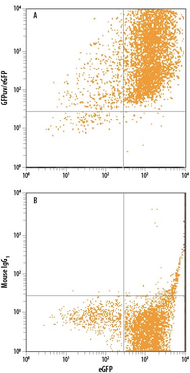
Detection of GFP in HEK293 Human Cell Line Transfected with eGFP by Flow Cytometry.
HEK293 human embryonic kidney cell line transfected with eGFP was stained with either (A) Mouse Anti-GFP PE-conjugated Monoclonal Antibody (Catalog # IC42401P) or (B) Mouse IgG1Phycoerythrin Isotype Control (Catalog # IC002P). To facilitate intracellular staining, cells were fixed with Flow Cytometry Fixation Buffer (Catalog # FC004) and permeabilized with Flow Cytometry Permeabilization/Wash Buffer I (Catalog # FC005). View our protocol for Staining Intracellular Molecules.Applications for GFP PE-conjugated Antibody
Intracellular Staining by Flow Cytometry
Sample: HEK293 human embryonic kidney cell line transfected with eGFP was fixed with Flow Cytometry Fixation Buffer (Catalog # FC004) and permeabilized with Flow Cytometry Permeabilization/Wash Buffer I (Catalog # FC005)
Formulation, Preparation, and Storage
Purification
Formulation
Shipping
Stability & Storage
- 12 months from date of receipt, 2 to 8 °C as supplied.
Background: GFP
Green fluorescent protein (GFP) is a 27 kDa protein originally isolated from the jellyfish Aequorea victoria. In the presence of UV light (490-520 nm), it emits a green fluorescent color that can be used to pinpoint locations of various intracellular proteins. GFP is 238 amino acids (aa) in length. It is a globular monomer that has a tendency to dimerize. The monomer has the shape of a beta-barrel with a chromophore (aa 65-67) containing alpha-helix running up its center. GFPuv is the Aequorea sequence with three aa substitutions; Phe to Ser at # 99, Met to Thr at # 153, and Val to Ala at # 163. This form expresses faster and is 18-fold brighter than native GFP; excitation peaks at 395 nm and emission at 508 nm.
Additional GFP Products
Product Documents for GFP PE-conjugated Antibody
Product Specific Notices for GFP PE-conjugated Antibody
For research use only