Histone H2AX [p Ser139] Antibody
Novus Biologicals, part of Bio-Techne | Catalog # NB100-2280

Key Product Details
Species Reactivity
Validated:
Human, Mouse, Canine
Cited:
Human, Mouse, Canine
Predicted:
Bat (100%), Bovine (100%), Chinese Hamster (100%), Guinea Pig (100%), Monkey (100%), Porcine (100%), Rabbit (100%). Backed by our 100% Guarantee.
Applications
Validated:
Immunocytochemistry/ Immunofluorescence, Immunohistochemistry, Immunohistochemistry-Frozen, Immunohistochemistry-Paraffin, Simple Western, Western Blot
Cited:
IF/IHC, Immunocytochemistry/ Immunofluorescence, Immunohistochemistry-Frozen, Immunohistochemistry-Paraffin, Western Blot
Label
Unconjugated
Antibody Source
Polyclonal Rabbit IgG
Concentration
0.1 mg/ml
Product Specifications
Immunogen
This Histone H2AX [p Ser139] Antibody was developed against a synthetic phospho-peptide, which represented a portion of the C-terminus of human histone H2AX surrounding phosphorylated serine 139 (GeneID 3014).
Reactivity Notes
Based on sequence percent identity: Gorilla (100%), Macaque (100%), Canine reactivity reported in scientific literature (PMID: 26991424).
Modification
p Ser139
Marker
DNA Double-strand break marker
Clonality
Polyclonal
Host
Rabbit
Isotype
IgG
Theoretical MW
15 kDa.
Disclaimer note: The observed molecular weight of the protein may vary from the listed predicted molecular weight due to post translational modifications, post translation cleavages, relative charges, and other experimental factors.
Disclaimer note: The observed molecular weight of the protein may vary from the listed predicted molecular weight due to post translational modifications, post translation cleavages, relative charges, and other experimental factors.
Scientific Data Images for Histone H2AX [p Ser139] Antibody
Immunohistochemistry: Histone H2AX [p Ser139] Antibody [NB100-2280] - Biological and molecular analysis of cutaneous wound healing in p73+/+ and p73-/- mice. Representative micrographs of immunohistochemistry (IHC) staining for Histone H2AX [p Ser139] in skin specimens from p73+/+ and p73-/- mice 10 days after wounding. All scale bars represent 50 um. Regions of the skin are labeled as: IFE, HF, epidermal wound edge (WE), and newly-formed epidermis of the wound (W); and the dotted line indicates the border between the WE and W. *p-value < 0.05, **p-value < 0.01, ***p-value < 0.001. Image collected and cropped by CiteAb from the following publication (https://dx.plos.org/10.1371/journal.pone.0218458), licensed under a CC-BY license.
Simple Western: Histone H2AX [p Ser139] Antibody [NB100-2280] - Simple Western lane view shows a specific band for Histone H2AX [p Ser139] in 0.2 mg/ml of Jurkat lysate(s). This experiment was performed under reducing conditions using the 12 - 230 kDa separation system.
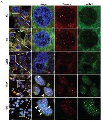
Immunohistochemistry: Histone H2AX [p Ser139] Antibody [NB100-2280] - Telomere dysfunctional induced foci (TIF) in HBV-related multistep hepatocarcinogenesis and the correlations thereof with stathmin and elongation factor 1alpha (EF1alpha) expression. A. Representative features of colocalization of Histone H2AX [p Ser139] and telomeric DNA in defined lesions of human multistep hepatocarcinogenesis. TIF are indicated by colored arrowheads: blue, DAPI; green, gamma H2AX; red, telomeres; yellow, TIF. Image collected and cropped by CiteAb from the following publication (https://translational-medicine.biomedcentral.com/articles/10.1186/1479-5876-12-154) licensed under a CC-BY license.
Applications for Histone H2AX [p Ser139] Antibody
Application
Recommended Usage
Immunocytochemistry/ Immunofluorescence
1:100 - 1:500
Immunohistochemistry-Paraffin
1:100-1:500
Simple Western
1:20
Western Blot
1:100-1:2000
Application Notes
Epitope exposure is recommended. Epitope exposure with citrate buffer will enhance staining. Likely to work with frozen sections. Use in WB reported in scientific literature ( PMID 24415760). Use in IHC-Frozen reported in scientific literature (PMID 26577699).
In Simple Western only 10 - 15 uL of the recommended dilution is used per data point.
See Simple Western Antibody Database for Simple Western validation: Tested in Jurkat lysate, separated by Size, antibody dilution of 1:20, apparent MW was 30 kDa. Separated by Size-Wes, Sally Sue/Peggy Sue.
In Simple Western only 10 - 15 uL of the recommended dilution is used per data point.
See Simple Western Antibody Database for Simple Western validation: Tested in Jurkat lysate, separated by Size, antibody dilution of 1:20, apparent MW was 30 kDa. Separated by Size-Wes, Sally Sue/Peggy Sue.
Reviewed Applications
Read 1 review rated 5 using NB100-2280 in the following applications:
Formulation, Preparation, and Storage
Purification
Immunogen affinity purified
Formulation
TBS and 0.1% BSA
Preservative
0.09% Sodium Azide
Concentration
0.1 mg/ml
Shipping
The product is shipped with polar packs. Upon receipt, store it immediately at the temperature recommended below.
Stability & Storage
Store at 4C. Do not freeze.
Background: Histone H2AX
References
1. Palla, V. V., Karaolanis, G., Katafigiotis, I., Anastasiou, I., Patapis, P., Dimitroulis, D., & Perrea, D. (2017). gamma-H2AX: Can it be established as a classical cancer prognostic factor?. Tumour biology : the journal of the International Society for Oncodevelopmental Biology and Medicine. https://doi.org/10.1177/1010428317695931
2. Kuo, L. J., & Yang, L. X. (2008). Gamma-H2AX - a novel biomarker for DNA double-strand breaks. In vivo (Athens, Greece).
3. Kinner, A., Wu, W., Staudt, C., & Iliakis, G. (2008). Gamma-H2AX in recognition and signaling of DNA double-strand breaks in the context of chromatin. Nucleic acids research. https://doi.org/10.1093/nar/gkn550
4. Redon, C. E., Weyemi, U., Parekh, P. R., Huang, D., Burrell, A. S., & Bonner, W. M. (2012). gamma-H2AX and other histone post-translational modifications in the clinic. Biochimica et biophysica acta. https://doi.org/10.1016/j.bbagrm.2012.02.021
5. H2AX: Uniprot (P16104)
Additional Histone H2AX Products
Product Documents for Histone H2AX [p Ser139] Antibody
Product Specific Notices for Histone H2AX [p Ser139] Antibody
Licensed to Novus Biologicals LLC under U.S. Patent Nos. 6,362,317 and 6,884,873.
This product is for research use only and is not approved for use in humans or in clinical diagnosis. Primary Antibodies are guaranteed for 1 year from date of receipt.
Loading...
Loading...
Loading...
Loading...
Loading...


![Immunocytochemistry/ Immunofluorescence: Histone H2AX [p Ser139] Antibody [NB100-2280] - Histone H2AX [p Ser139] Antibody](https://resources.bio-techne.com/images/products/nb100-2280_rabbit-polyclonal-histone-h2ax-p-ser139-antibody-310202415175243.jpg)
![Immunocytochemistry/ Immunofluorescence: Histone H2AX [p Ser139] Antibody [NB100-2280] - Histone H2AX [p Ser139] Antibody](https://resources.bio-techne.com/images/products/nb100-2280_rabbit-polyclonal-histone-h2ax-p-ser139-antibody-310202415171325.jpg)
![Immunocytochemistry/ Immunofluorescence: Histone H2AX [p Ser139] Antibody [NB100-2280] - Histone H2AX [p Ser139] Antibody](https://resources.bio-techne.com/images/products/nb100-2280_rabbit-polyclonal-histone-h2ax-p-ser139-antibody-3102024165725.jpg)
![Immunohistochemistry: Histone H2AX [p Ser139] Antibody [NB100-2280] - Histone H2AX [p Ser139] Antibody](https://resources.bio-techne.com/images/products/nb100-2280_rabbit-polyclonal-histone-h2ax-p-ser139-antibody-310202415541940.jpg)
![Western Blot: Histone H2AX [p Ser139] Antibody [NB100-2280] - Histone H2AX [p Ser139] Antibody](https://resources.bio-techne.com/images/products/nb100-2280_rabbit-polyclonal-histone-h2ax-p-ser139-antibody-31020241554196.jpg)
![Immunocytochemistry/ Immunofluorescence: Histone H2AX [p Ser139] Antibody [NB100-2280] - Histone H2AX [p Ser139] Antibody](https://resources.bio-techne.com/images/products/nb100-2280_rabbit-polyclonal-histone-h2ax-p-ser139-antibody-3102024165746.jpg)
![Immunohistochemistry: Histone H2AX [p Ser139] Antibody [NB100-2280] - Histone H2AX [p Ser139] Antibody](https://resources.bio-techne.com/images/products/nb100-2280_rabbit-polyclonal-histone-h2ax-p-ser139-antibody-31020241612641.jpg)
![Immunocytochemistry/ Immunofluorescence: Histone H2AX [p Ser139] Antibody [NB100-2280] - Histone H2AX [p Ser139] Antibody](https://resources.bio-techne.com/images/products/nb100-2280_rabbit-polyclonal-histone-h2ax-p-ser139-antibody-31020241612651.jpg)