Human 4-1BB/TNFRSF9/CD137 Antibody
R&D Systems, part of Bio-Techne | Catalog # MAB8381
Recombinant Monoclonal Antibody.


Conjugate
Catalog #
Key Product Details
Validated by
Biological Validation
Species Reactivity
Human
Applications
CyTOF-ready, Flow Cytometry, Immunocytochemistry, Simple Western, Western Blot
Label
Unconjugated
Antibody Source
Recombinant Monoclonal Rabbit IgG Clone # 2356B
Product Specifications
Immunogen
Chinese hamster ovary cell line CHO-derived recombinant human 4‑1BB/TNFRSF9/CD137
Leu24-His183
Accession # Q07011
Leu24-His183
Accession # Q07011
Specificity
Detects human 4‑1BB/TNFRSF9/CD137 in direct ELISAs.
Clonality
Monoclonal
Host
Rabbit
Isotype
IgG
Scientific Data Images for Human 4-1BB/TNFRSF9/CD137 Antibody
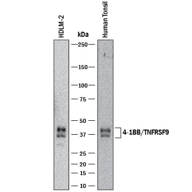
Detection of Human 4‑1BB/TNFRSF9/CD137 by Western Blot.
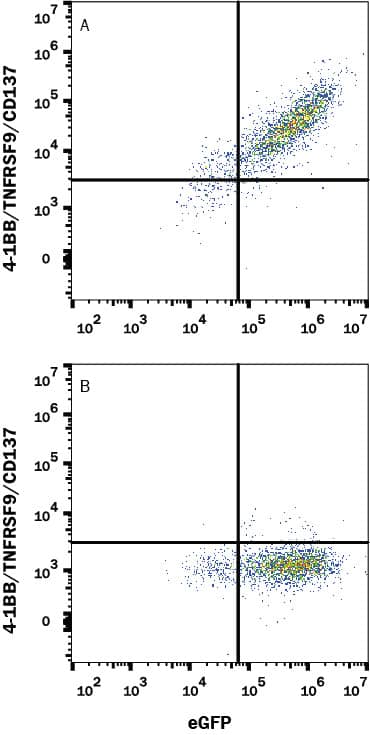
Western blot shows lysates of HDLM-2 human Hodgkin's lymphoma cell line and human tonsil tissue. PVDF membrane was probed with 2 µg/mL of Rabbit Anti-Human 4-1BB/TNFRSF9/CD137 Monoclonal Antibody (Catalog # MAB8381) followed by HRP-conjugated Anti-Rabbit IgG Secondary Antibody (HAF008). Specific bands were detected for 4-1BB/TNFRSF9/CD137 at approximately 32 and 40 kDa (as indicated). This experiment was conducted under reducing conditions and using Immunoblot Buffer Group 1.Detection of 4-1BB/TNFRSF9/CD137 in HEK293 Human Cell Line Transfected with Human 4-1BB/TNFRSF9/CD137 and eGFP by Flow Cytometry.
HEK293 human embryonic kidney cell line transfected with either (A) human 4-1BB/TNFRSF9/CD137 or (B) irrelevant protein, and eGFP was stained with Rabbit Anti-Human 4-1BB/TNFRSF9/CD137 Monoclonal Antibody (Catalog # MAB8381) followed by APC-conjugated Goat-anti Rabbit IgG secondary antibody (F0111). Quadrant markers were set based on control antibody staining (MAB1050). View our protocol for Staining Membrane-associated Proteins.4‑1BB/TNFRSF9/CD137 in Human PBMCs and HEK293 Cell Line.
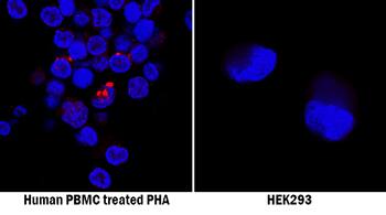
4-1BB/TNFRSF9/CD137 was detected in immersion fixed peripheral blood mononuclear cells (PBMCs) treated with PHA (positive control, left panel) and HEK293 human embryonic kidney cell line (negative control, right panel) using Rabbit Anti-Human 4-1BB/TNFRSF9/CD137 Monoclonal Antibody (Catalog # MAB8381) at 8 µg/mL for 3 hours at room temperature. Cells were stained using the NorthernLights™ 557-conjugated Anti-Rabbit IgG Secondary Antibody (red NL004) and counterstained with DAPI (blue). Specific staining was localized to cell surfaces. View our protocol for Fluorescent ICC Staining of Non-adherent Cells.Applications for Human 4-1BB/TNFRSF9/CD137 Antibody
Application
Recommended Usage
CyTOF-ready
Ready to be labeled using established conjugation methods. No BSA or other carrier proteins that could interfere with conjugation.
Flow Cytometry
0.25 µg/106 cells
Sample: HEK293 Human Cell Line Transfected with Human 4-1BB/TNFRSF9/CD137 and eGFP
Sample: HEK293 Human Cell Line Transfected with Human 4-1BB/TNFRSF9/CD137 and eGFP
Immunocytochemistry
8-25 µg/mL
Sample: Immersion fixed human peripheral blood mononuclear cells (PBMCs) and HEK293 human embryonic kidney cell line
Sample: Immersion fixed human peripheral blood mononuclear cells (PBMCs) and HEK293 human embryonic kidney cell line
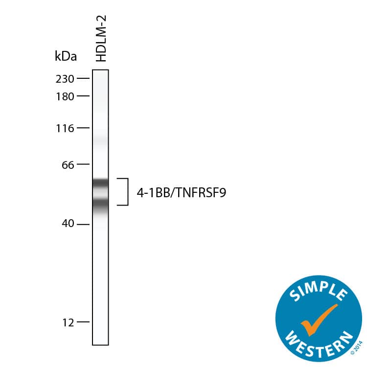
Simple Western
20 µg/mL
Sample: HDLM-2 human Hodgkin's lymphoma cells
Sample: HDLM-2 human Hodgkin's lymphoma cells
Western Blot
2 µg/mL
Sample: HDLM‑2 human Hodgkin's lymphoma cell line and human tonsil tissue
Sample: HDLM‑2 human Hodgkin's lymphoma cell line and human tonsil tissue
Formulation, Preparation, and Storage
Purification
Protein A or G purified from cell culture supernatant
Reconstitution
Reconstitute at 0.5 mg/mL in sterile PBS. For liquid material, refer to CoA for concentration.
Formulation
Lyophilized from a 0.2 μm filtered solution in PBS with Trehalose. *Small pack size (SP) is supplied either lyophilized or as a 0.2 µm filtered solution in PBS.
Shipping
Lyophilized product is shipped at ambient temperature. Liquid small pack size (-SP) is shipped with polar packs. Upon receipt, store immediately at the temperature recommended below.
Stability & Storage
Use a manual defrost freezer and avoid repeated freeze-thaw cycles.
- 12 months from date of receipt, -20 to -70 °C as supplied.
- 1 month, 2 to 8 °C under sterile conditions after reconstitution.
- 6 months, -20 to -70 °C under sterile conditions after reconstitution.
Background: 4-1BB/TNFRSF9/CD137
4-1BB activation enhances CD8+ T cell and NK cell mediated anti-tumor immunity (14). It also contributes to the development of inflammation in high fat diet-induced metabolic syndrome (15). Soluble forms of 4-1BB and 4-1BB Ligand circulate at elevated levels in the serum of rheumatoid arthritis and hematologic cancer patients, respectively (16, 17).
References
- Wang, C. et al. (2009) Immunol. Rev. 229:192.
- Schwarz, H. et al. (1993) Gene 134:295.
- Alderson, M.R. et al. (1994) Eur. J. Immunol. 24:2219.
- Wen, T. et al. (2002) J. Immunol. 168:4897.
- Pulle, G. et al. (2006) J. Immunol. 176:2739.
- Zheng, G. et al. (2004) J. Immunol. 173:2428.
- Kim, D. et al. (2008) J. Immunol. 180:2062.
- Lee, S. et al. (2008) Nat. Immunol. 9:917.
- Choi, B.K. et al. (2009) J. Immunol. 182:4107.
- Nishimoto, H. et al. (2005) Blood 106:4241.
- Saito, K. et al. (2004) J. Biol. Chem. 279:13555.
- Lee, S. et al. (2005) J. Immunol. 174:6803.
- Ma, B.Y. et al. (2005) Blood 106:2002.
- Choi, B.K. et al. (2010) J. Immunol. 185:1404.
- Kim, C. et al. (2011) Diabetes 60:3159.
- Michel, J. et al. (1998) Eur. J. Immunol. 28:290.
- Salih, H.R. et al. (2001) J. Immunol. 167:4059.
Alternate Names
41BB, CD137, ILA, TNFRSF9
Gene Symbol
TNFRSF9
UniProt
Additional 4-1BB/TNFRSF9/CD137 Products
Product Documents for Human 4-1BB/TNFRSF9/CD137 Antibody
Product Specific Notices for Human 4-1BB/TNFRSF9/CD137 Antibody
For research use only
Loading...
Loading...
Loading...
Loading...
Loading...


