Human ALK-1 Antibody
R&D Systems, part of Bio-Techne | Catalog # MAB370

Key Product Details
Species Reactivity
Validated:
Cited:
Applications
Validated:
Cited:
Label
Antibody Source
Product Specifications
Immunogen
Asp22-Gln118
Accession # P37023
Specificity
Clonality
Host
Isotype
Scientific Data Images for Human ALK-1 Antibody
Detection of Human ALK-1 by Western Blot.
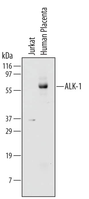
Western blot shows lysates of Jurkat human acute T cell leukemia cell line and human placenta tissue. PVDF membrane was probed with 2 µg/mL of Mouse Anti-Human ALK-1 Monoclonal Antibody (Catalog # MAB370) followed by HRP-conjugated Anti-Mouse IgG Secondary Antibody (Catalog # HAF007). A specific band was detected for ALK-1 at approximately 65 kDa (as indicated). This experiment was conducted under reducing conditions and using Immunoblot Buffer Group 1.Detection of Human ALK‑1 by Simple WesternTM.
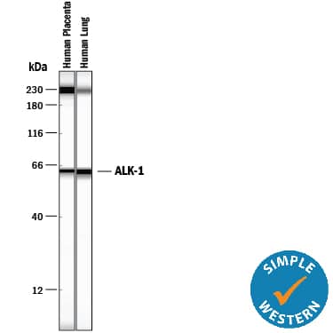
Simple Western lane view shows lysates of human placenta tissue and human lung tissue, loaded at 0.2 mg/mL. A specific band was detected for ALK‑1 at approximately 63 kDa (as indicated) using 40 µg/mL of Mouse Anti-Human ALK‑1 Monoclonal Antibody (Catalog # MAB370). This experiment was conducted under reducing conditions and using the 12-230 kDa separation system.Non-specific interaction with the 230 kDa Simple Western standard may be seen with this antibody.Applications for Human ALK-1 Antibody
Simple Western
Sample: Human placenta tissue and human lung tissue
Western Blot
Sample: Human placenta tissue
Formulation, Preparation, and Storage
Purification
Reconstitution
Formulation
Shipping
Stability & Storage
- 12 months from date of receipt, -20 to -70 °C as supplied.
- 1 month, 2 to 8 °C under sterile conditions after reconstitution.
- 6 months, -20 to -70 °C under sterile conditions after reconstitution.
Background: ALK-1
Transforming Growth Factor beta (TGF-beta) superfamily ligands exert their biological activities via binding to heteromeric receptor complexes of two types (I and II) of serine/threonine kinases. Type II receptors are constitutively active kinases that phosphorylate type I receptors upon ligand binding. In turn, activated type I kinases phosphorylate downstream signaling molecules including the various smads. Transmembrane proteoglycans, including the type III receptor (betaglycan) and endoglin, can bind and present some of the TGF-beta superfamily ligands to type I and II receptor complexes and enhance their cellular responses. Seven type I receptors (also termed activin receptor-like kinase (ALK)) and five type II receptors have been isolated from mammals. ALK-2, -3, -4, -5, and -6 are also known as Activin R1A, BMPR-1A, Activin R1B, TGF-beta R1, and BMPR-1B, respectively, reflecting their ligand preferences. Evidence suggests that TGF-beta 1, TGF-beta 3 and an unknown ligand present in serum can activate chimeric ALK-1. ALK-1 shares with other type I receptors a cysteine-rich domain with conserved cysteine spacing in the extracellular region, and a glycine- and serine-rich domain (the GS domain) preceding the kinase domain. ALK-1 is expressed highly in endothelial cells and other highly vascularized tissues. The expression patterns of ALK-1 parallels that of endoglin. Mutations in ALK-1 as well as in endoglin are associated with hereditary hemorrhagic telangiectasia (HHT), suggesting a critical role for ALK-1 in the control of blood vessel development or repair. Human and mouse ALK-1 share approximately 71% amino acid sequence identity in their extracellular regions.
References
- ten Dijke, P. et al. (1993) Oncogene 8:2879.
- ten Dijke, P. et al. (1994) Science 264:101.
- Lux, A. et al. (1999) J. Biol. Chem. 274:9984.
Long Name
Alternate Names
Gene Symbol
UniProt
Additional ALK-1 Products
Product Documents for Human ALK-1 Antibody
Product Specific Notices for Human ALK-1 Antibody
For research use only

