Human Cathepsin B Antibody
R&D Systems, part of Bio-Techne | Catalog # AF953

Key Product Details
Validated by
Species Reactivity
Validated:
Cited:
Applications
Validated:
Cited:
Label
Antibody Source
Product Specifications
Immunogen
Arg18-Ile339
Accession # P07858
Specificity
Clonality
Host
Isotype
Scientific Data Images for Human Cathepsin B Antibody
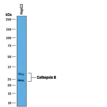
Detection of Human Cathepsin B by Western Blot.
Western blot shows lysates of HepG2 human hepatocellular carcinoma cell line. PVDF membrane was probed with 0.25 µg/mL of Goat Anti-Human Cathepsin B Antigen Affinity-purified Polyclonal Antibody (Catalog # AF953) followed by HRP-conjugated Anti-Goat IgG Secondary Antibody (Catalog # HAF019). A specific band was detected for Cathepsin B at approximately 25-30 kDa (as indicated). This experiment was conducted under reducing conditions and using Immunoblot Buffer Group 1.Cathepsin B in Human Brain.
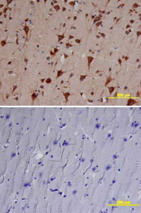
Cathepsin B was detected in immersion fixed paraffin-embedded sections of human brain (cortex) using Goat Anti-Human Cathepsin B Antigen Affinity-purified Polyclonal Antibody (Catalog # AF953) at 10 µg/mL overnight at 4 °C. Tissue was stained using the Anti-Goat HRP-DAB Cell & Tissue Staining Kit (brown; CTS008) and counterstained with hematoxylin (blue). Lower panel shows a lack of labeling if primary antibodies are omitted and tissue is stained only with secondary antibody followed by incubation with detection reagents. View our protocol for Chromogenic IHC Staining of Paraffin-embedded Tissue Sections.Detection of Human Cathepsin B by Simple WesternTM.
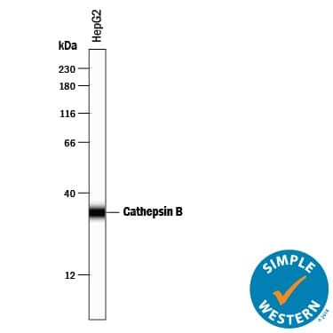
Simple Western lane view shows lysates of HepG2 human hepatocellular carcinoma cell line, loaded at 0.2 mg/mL. A specific band was detected for Cathepsin B at approximately 34 kDa (as indicated) using 2.5 µg/mL of Goat Anti-Human Cathepsin B Antigen Affinity-purified Polyclonal Antibody (Catalog # AF953) followed by 1:50 dilution of HRP-conjugated Anti-Goat IgG Secondary Antibody (HAF109). This experiment was conducted under reducing conditions and using the 12-230 kDa separation system.Applications for Human Cathepsin B Antibody
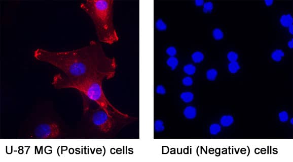
Immunocytochemistry
Sample: Immersion fixed U‑87 MG human glioblastoma/astrocytoma cell line (Positive) & Daudi human Burkitt's lymphoma cell line (Negative)
Immunohistochemistry
Sample: Immersion fixed paraffin-embedded sections of human normal brain (cortex) and bladder cancer tissue
Immunoprecipitation
Sample: Conditioned cell culture medium spiked with Recombinant Human Cathepsin B (Catalog # 953-CY), see our available Western blot detection antibodies
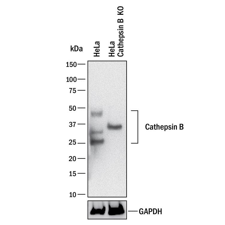
Knockout Validated
Simple Western
Sample: HepG2 human hepatocellular carcinoma cell line
Western Blot
Sample: HepG2 human hepatocellular carcinoma cell line
Reviewed Applications
Read 3 reviews rated 5 using AF953 in the following applications:
Formulation, Preparation, and Storage
Purification
Reconstitution
Formulation
Shipping
Stability & Storage
- 12 months from date of receipt, -20 to -70 °C as supplied.
- 1 month, 2 to 8 °C under sterile conditions after reconstitution.
- 6 months, -20 to -70 °C under sterile conditions after reconstitution.
Background: Cathepsin B
Cathepsin B is the first described member of the family of lysosomal cysteine proteases (1). Cathepsin B possesses both endopeptidase and exopeptidase activities, in the latter case acting as a peptidyl-dipeptidase. It is known to process a number of proteins, including pro and active caspases, prorenin, and secretory leucoprotease inhibitor (SLPI) (2-4). Therefore, Cathepsin B may play a role in activation and inactivation of caspases, activation of renin and inactivation of SLPI, the key steps in apoptosis, angiotensin production, and progression of emphysema, respectively. Because of its increased levels and redistribution of the enzyme in human and animal tumors, Cathepsin B may also have role in invasion and metastasis (5).
In addition to lysosome, Cathepsin B can be secreted or associated with plasma membrane, cytoplasm, and nucleus. It is synthesized as a preproenzyme. Following removal of the signal peptide, the inactive proenzyme undergoes further modifications including removal of the pro region to result in the active enzyme (1).
References
- Mort, J.S. (2004) in Handbook of Proteolytic Enzymes. Barrett, A.J. et al. (eds): Academic Press, San Diego, p. 1079.
- Vancompernolle, K. et al. (1998) FEBS Lett. 438:150.
- Jutras, I. and T.L. Reudelhuber (1999) FEBS Lett. 443:48.
- Taggart, C.C. et al. (2001) J. Biol. Chem. 276:33345.
- Bergquin, I.M. and B.F. Sloane (1996) Adv. Exp. Med. Biol. 389:281.
Alternate Names
Gene Symbol
UniProt
Additional Cathepsin B Products
Product Documents for Human Cathepsin B Antibody
Product Specific Notices for Human Cathepsin B Antibody
For research use only
